

A cataract is any opacification of the normally clear lens of the eye. The lens sits just behind the iris and is viewable through the pupil. Cataracts can develop in both old dogs (this is called senile cataracts) and young dogs (juvenile cataracts, congenital cataracts). They can develop in one eye, or both. They may develop slowly, or happen all of a sudden. Sometimes they are obvious and sometimes they are difficult to see, even for your veterinarian.
Symptoms of Cataracts in Progressively Developing Stages
Cataracts are identified by their level of development:
Stage 1 – Incipient cataracts
These are the smallest, earliest cataracts and are difficult to impossible to see with the naked eye – and sometimes even hard for your veterinarian to identify without special equipment. Incipient cataracts affect less than 15% of the lens. Sometimes the only sign of incipient cataracts is that the dog can’t seem to catch treats or balls thrown to him like he used to.
Stage 2 – Immature cataracts
These are easier to see but difficult to differentiate from lenticular or nuclear sclerosis, a normal aging cloudiness of the lens that will never result in complete blindness like cataracts do. Immature cataracts look hazy, grayish-blue. They may involve only a small portion of the lens or the entire lens. They are opaque (in contrast to nuclear sclerosis, which is merely translucent), but the density of the opacity is low enough to allow some light to get through to the retina. These dogs will likely be able to see well enough to navigate around the house without trouble, but they will not see well enough to catch treats or balls, nor to see your facial expressions, small hand movements, etc.
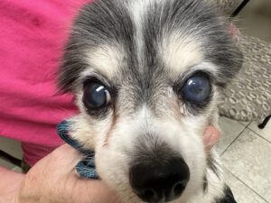
Stage 3 – Mature cataracts
These are impossible to miss. Mature cataracts in dogs involve the entire lens and are densely opaque, looking like a white crystal filling the entire pupil. If only one eye is affected, the dog will still be happily visual. Mature cataracts in both eyes make the dog completely blind. No light can get through these extremely dense, opacified lenses.
Stage 4 – Hypermature cataracts
This is the final stage, and the worst, as it is most likely to cause painful secondary issues within the eye. Not all cataracts will progress to the hypermature stage. The ones that do begin to shrink as they degenerate, resulting in a change in the size and/or shape of the cataract. They may leak proteinaceous fluid as the lens liquefies. The fluid released often results in severe, painful inflammation within the eye called lens-induced uveitis, which can then lead to glaucoma.
Signs of lens-induced uveitis include:
- Painful eye
- Red eye
- Engorged blood vessels on the white of the eye
- Corneal edema (bluish haze of the normally clear cornea)
- Blood or pus-like material within the eye (between the cornea and the iris)
- Swelling, mottling or change in color of the iris
- Contracted pupil
What hypermature cataracts look like varies tremendously. They may look simply like smaller mature cataracts (the dog may even regain some vision around the edges as the cataract shrinks), the crystalline appearance may sink to only the bottom half of the lens capsule, or you may see almost like a wrinkled appearance to the cataract.
Causes of canine cataracts
The lens of the eye is made up of protein and water. Because it’s protein, the lens can be damaged, similar to any other tissue made of protein in the body, such as muscle tissue. This means that trauma to the eye (penetrating or blunt force) and/or chronic inflammation in the eye can result in cataract formation.
Cataracts can be inherited, with a genetic predisposition in certain breeds, including the Staffordshire Terrier, Boston Terrier, Australian Shepherd, and French Bulldog. Affected dogs should not be used for breeding. Genetic testing is available through the Veterinary Genetics Laboratory at the University of California, Davis, School of Veterinary Medicine.
A nutritional cause for cataracts has been identified in orphan puppies who were fed milk replacer.
Diabetes, though, may be the most common cause of cataracts in dogs. The excess sugar in diabetic dogs’ bloodstream is converted to sorbitol in the lens. The sorbitol draws water into the lens, which causes the lens to swell. This creates oxidative stress and disrupts the fibers in the lens, resulting in cataract formation. About 75-80% of dogs with diabetes will develop cataracts within the first year of their diagnosis, regardless of how well controlled their diabetes is. Diabetic cataracts tend to form quickly and frequently cause secondary issues, including lens-induced uveitis and glaucoma (increased intraocular pressure). Glaucoma in diabetic dogs is notoriously difficult to manage and often ends with surgical removal of the painful, blind eye (enucleation). Your best bet for a diabetic dog with cataracts is surgical correction as soon as possible.
Surgical Treatment for Cataracts in Dogs
If your dog is diagnosed with cataracts, the best treatment – the gold standard of care – is having cataract surgery as soon as possible. The best outcomes with the fewest complications are achieved when surgery is performed before the cataracts become mature. Time is of the essence: If too much time passes before cataracts are diagnosed and treated with surgery, some cataracts will develop characteristics that preclude surgery from being performed at all.
As long as the dog’s retina and the rest of the eye is healthy, and the dog’s general health is good enough to withstand anesthesia, surgery is warranted.
Prior to surgery an electroretinogram will be performed to ensure that the dog has healthy retinas and will indeed be able to see after surgery. The surgery, which is performed by a board-certified veterinary ophthalmologist, is called phacoemulsification. Small incisions are made in the cornea and the lens capsule. High-frequency vibration is used to essentially pulverize the lens, which is then removed by vacuum. An artificial lens is inserted into the eye and the cornea is sutured closed. Most dogs spend three to four days in the hospital receiving critical post-op treatments and careful monitoring. At home, the dog will require continued treatment with several eye drops administered four to six times a day for several weeks.
Follow-up exams with the ophthalmologist are important to check for and treat any post-operative complications that arise. The most common post-operative complications are prolonged intraocular inflammation and glaucoma. Again, the more mature the cataract before surgery is performed, the more likely post-operative complications are.
When Cataract Surgery Is Contraindicated
Are there reasons NOT to pursue cataract surgery? There sure are. These include:
- General health of the dog. Is the dog a good candidate for general anesthesia? Not always. The risk of the general anesthesia required for the procedure may be too great, especially in older dogs with heart and/or kidney issues. Does the dog have a reasonable life expectancy after surgery? This depends on the dog’s age and other factors like cancer status.
- Condition of the eyes. Are the corneas healthy? This is important for a good surgical outcome and also for vision. Are the retinas healthy? If not, the dog will still be blind, or will become blind, in spite of surgery.
- Ability of the dog owner to commit. Post-operative care after cataract surgery is pretty intense, with topical medications required to be administered frequently around the clock. Sometimes lifestyles or work schedules can get in the way of this.
- Temperament of the dog. Let’s face it. Some dogs are going to be impossible to medicate/manage with such an intense post-operative protocol, both in the hospital and at home afterward. This is unfortunately something that must be seriously considered.
Non-Surgical Care for Cataracts
What if your dog is not a good candidate for cataract surgery?
Your veterinarian can prescribe anti-inflammatory eye drops to help prevent cataract-associated inflammatory ocular disease. Additionally, it is recommended you have your dog’s eye pressures checked for glaucoma every four to six months.
All dogs benefit from being maintained at a healthy weight, but obesity is more consequential for dogs with cataracts, because obesity predisposes dogs to diabetes and diabetes causes cataracts. Keep your dog slim!
Owners should be alert to any sign that their dog is experiencing eye pain, such as squinting, sensitivity to light, rubbing at the eye, lethargy, and inappetence. Make an appointment with your veterinarian if these signs are observed.
Finally, if your dog becomes blind from her cataracts, do everything you can to make life as easy as possible for her. This article has many helpful tips, including a recommendation to read the book, Living With Blind Dogs: A Resource Book and Training Guide for the Owners of Blind and Low-Vision Dogs, by Caroline D. Levin.
To tell the difference between cataracts and other causes of cloudy eyes in dogs, we must distinguish whether the cloudiness is deep inside the eye behind the pupil, or on the outer surface of the eye or cornea.
Ocular diseases that can cause cloudy corneas in dogs include:
- Glaucoma (increased intraocular pressure)
- Corneal dystrophy (abnormalities of layers in the cornea resulting in corneal opacities)
- Corneal ulcer (scratch or abrasion disrupting the integrity of the cornea)
- Uveitis (intraocular inflammation of the middle layers of the eye including the iris)
- Keratitis (inflammation of the cornea)
- Dry Eye Syndrome (also known as keratoconjunctivitis sicca or KCS, decreased tear production resulting in corneal irritation)
- Corneal edema (fluid accumulation within the layers of the cornea)
Cloudiness behind the pupil localizes the lesion to the lens, making the cause either cataracts or nuclear sclerosis. Immature cataracts and nuclear sclerosis can be difficult to differentiate, as they both tend to look hazy and grayish-bluish in color. Your veterinarian may be able to distinguish between the two, or can refer you to a veterinary ophthalmologist for specialized assessment.