


There are many possible causes of dental disease in dogs including plaque accumulation, tooth trauma, genetic predispositions, cavities, and cancerous conditions. In recent years, there has been an increase in the research of canine tooth root resorption – a process in which part or parts of the tooth are destroyed by physiologic processes gone awry – as a cause of dental disease.
Unfortunately, the research has not yet resulted in solid answers as to the cause or effective treatment of tooth root resorption in dogs. Nevertheless, owners should be aware of the condition so they can bring any abnormality they may observe in their dog’s mouth to their veterinarian’s attention.
ANATOMY OF A TOOTH PROBLEM
A healthy canine tooth consists of the crown – the part of the tooth above the gumline – and the root, which is below the gumline and makes up the majority of the tooth.
The center of the tooth root is the pulp chamber; this is living tissue filled with nerve endings and blood vessels. Moving outward from the core of the tooth, the next layer is dentin, a hard, bone-like material that protects the pulp. Outside of the dentin, cementum is a thin coating that attaches the tooth to the jaw bone.
Enamel, the white, visible part of the tooth, protects the crown. It is the hardest substance in the body. Once laid down, enamel is no longer produced. Any destruction to enamel is permanent.
Tooth root resorption occurs when a part or parts of the tooth are destroyed. Any area of the tooth can be affected, from the dentin to the enamel.
Odontoclasts are cells that are critical for resorbing tooth tissue. They are usually associated with breakdown of baby tooth roots, so that these teeth can fall out and make way for permanent teeth. For reasons that are not understood, sometimes the supportive “net” of proteins that protect the teeth gets compromised, and odontoclasts can then break down the tooth and surrounding tissues (called the periodontium) without restriction.
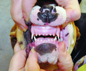
The beginning of this breakdown appears as a lesion that looks like a pit or a hole in the tooth. These lesions are often found at the junction where the enamel meets the cementum. If above the gumline, bright pink tissue from the gums (called gingiva) may cover the hole. This is the body’s way of trying to heal the defect. If the dog is under anesthesia for a dental cleaning, once the tooth is scraped clean, the lesion becomes obvious. However, many of the lesions are below the gumline and will not be seen except through dental x-rays.
There are three basic types of resorptive lesions: physiologic, inflammatory, and non-inflammatory.
Physiologic resorption is normal and occurs as the body prepares to shed the deciduous teeth (baby teeth) as the adult teeth develop and emerge.
Inflammatory and non-inflammatory resorptive lesions are abnormal. The triggers for these types of tooth destruction are unknown. One study found a possible correlation between age and breed, with older, large breed dogs predisposed.
SIGNS
What symptoms might your dog show? This is what makes resorption lesions confusing! There may be no symptoms at all. Resorptive lesions are often found incidentally if your dog has full mouth x-rays before a dental cleaning. (This is one reason that x-rays are such an important part of dental cleaning.) If symptoms are noted, they might include drooling, difficulty chewing, bad breath, mouth pain, and dropping food when chewing.
If a resorptive lesion affects the crown of the tooth, a hole may be seen. It is also possible to see pink fleshy tissue that appears to be growing on the tooth. This is gingival tissue that is trying to cover the defect in the crown. It is a hallmark of a resorptive lesion.
DO YOU HAVE TO DO ANYTHING ABOUT THIS?
Treatment for this condition remains controversial. In some cases, your veterinarian will suggest only monitoring every six months with dental x-rays. But if discomfort or dysfunction are observed, extraction of the affected tooth is indicated.
In the past, veterinary dental specialists tried filling these resorptive lesions like a dentist would fill a cavity in a human. However, this proved to be a mostly unsuccessful tack; usually, the tooth loss would continue to progress. The filling would fall out and the tooth would have to be extracted after all. If the dog seems to be experiencing discomfort, and/or the loss of the tooth is certain, the tooth should just be extracted.
Unfortunately, there is no known way to prevent tooth resorption, as the causes are not known. However, dogs who have suffered one tooth resorption are prone to more. As a result, close attention to overall dental health is critical throughout your dog’s life. If your veterinarian suspects resorptive lesions, then referral to a veterinary dentist may be the best course of action. Canine resorptive lesions are an area of limited knowledge and current research, so speaking with a specialist might be the best bet.
Catherine Ashe graduated the University of Tennessee College of Veterinary Medicine in 2008. After a small animal intensive emergency internship, she practiced ER medicine for nine years. She works as a relief veterinarian in Asheville, North Carolina and loves the general practice side of medicine.


My dog has black on the,top of her teeth what causes this
What is the symptoms for a dog tooth problem and how to help the dog feel better?