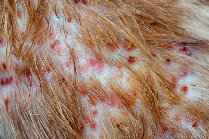
Folliculitis is inflammation of the hair follicles. Inflammation is the body’s response to an injury or infection of some sort; when hair follicles become infected with bacteria, the body sends inflammatory cells to attack the bacteria and heal damaged tissue. In the case of foliliculitis in dogs, the original insult may have been caused by ectoparasites, a fungus, an underlying endocrine disorder, or trauma caused by the scratching and chewing that results from the itching of hypersensitivity (allergy). The condition is sometimes called superficial pyoderma; superficial refers to the hair follicles and the epidermis; pyo means pus and derma means skin.
Folliculitis can occur anywhere on a dog’s body, but is found most frequently in warm, moist areas of the dog’s skin, particularly where there is a skin fold, such as the dog’s lips, face, neck, vulva, and the base of the tail. The axillary region (or armpits as we know them on human bodies), groin region, and spaces between the toes are also prime locations for developing folliculitis.

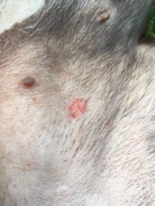
Bacterial folliculitis begins as small, flat red circles on your dog’s skin called macules. As the condition progresses, the macules become raised bumps called papules. Papules may fill with pus, creating a small white dot in the middle of the raised red circle. When pus fills a papule on a dog, it is called a pustule; in humans, we’d call this a pimple.
Folliculitis is pruritic (itchy) and you may notice your dog scratching or licking at these lesions. Scratching or licking at papules and pustules causes them to rupture and release clear fluid from papules and pus from pustules. When that clear fluid and pus dries, the papules and pustules become covered with a crust. If your dog continues licking and scratching at these crusty red circles, the circles get bigger and develop scales or flakes along the edge. These are called epidermal collarettes.
There are three types of pyoderma: surface, superficial, and deep.
- Surface pyoderma is an infection on the skin surface. “Hot spots” – those small, red, itchy patches that tend to appear on the neck, face, and rump near the tail during the hot, humid months of summer – are examples of surface pyoderma.
- Superficial pyoderma is an infection of the epidermis (the top layers of skin) and the hair follicles. Folliculitis is an infection of the hair follicles and the superficial skin.
- Deep pyoderma is an infection that extends into the dermis, or the deep layer of the skin.
General Treatment of Folliculitis in Dogs
Treatment of folliculitis may include a medicated shampoo to reduce the bacterial population on the skin and ease the pruritus and discomfort. Treatment will also likely include an oral antibiotic. A dog will typically need to be on an oral antibiotic until one week after all of his symptoms resolve. This may take as little as two weeks but usually requires four to six weeks of therapy.
Folliculitis in Dogs Causes and Targeted Treatment
A bacterial infection of the skin and hair follicles is almost always secondary to one of many potential problems with the skin. Successful treatment of folliculitis will depend on its original cause. These most common precipitating conditions include:
Demodicosis
This is caused by a mite called Demodex, which live in the hair follicles and sebaceous glands of your dog’s skin. They are a commensal mite; they live on and benefit from your dog without causing your dog harm. Demodex mites can cause a dog to become itchy when their populations suddenly increase. (When the equilibrium between the Demodex mites and the skin microenvironment becomes unbalanced, and the mite population explodes, they are then considered a parasite, rather than a commensal.)
Puppies are more prone to developing pruritus caused by the Demodex mite because of their young age. Adult dogs can also develop demodicosis but there is often an underlying immunocompromising condition that allows the Demodex mite to proliferate. Demodex mites are not contagious to other dogs.
Your veterinarian may want to complete a skin scrape test to look for Demodex mites. There are several treatments for demodicosis. The only FDA-approved medication for demodicosis is a dip treatment called amitraz (brand name Mitaban). There are side effects to using amitraz and the odor of the dip is quite noxious. Other treatments for demodicosis are not FDA-approved for this purpose but have shown good efficacy in treating the condition. These treatments include ivermectin (an oral medication), milbemycin (found in several heartworm preventatives), moxidectin (found in some topical flea preventatives), and the fluralaner class of drugs (found in several oral flea/tick preventatives). Discuss with your veterinarian which treatment option is best for you and your dog.
Ectoparasites (skin parasites)
Skin parasites include mites and lice that do not belong on your dog. They include:
- The Sarcoptes scabiei mite is the cause of sarcoptic mange, aka scabies. This mite burrows into your dog’s skin and lays eggs in the tunnels they create. Dogs have an allergic reaction to the poop that mites leave behind in the skin tunnels, causing the dog to become extremely itchy.
- Cheyletiella yasguri (the cause of cheyletiellosis) is a white mite that lives on the surface of a dog’s skin. Dogs infected with Cheyletiella have dandruff that looks like it is moving. This infection is often called “walking dandruff.”
- Trombiculidae, better known as “chiggers,” are tiny, orange-red mites that attach to a dog’s skin to feed for a few days before detaching.
The treatment for all three of these mites is similar and include amitraz and lime-sulfur topical dips, ivermectin (an oral medication), moxidectin and selamectin (found in some topical flea preventatives), and the fluralaner class of drugs (found in several oral flea/tick preventatives). Moxidectin and selamectin are FDA-approved for treating sarcoptes mange but have been found effective at treating walking dandruff and chiggers. The fluralaner class of drug is not FDA-approved for any of these mites but has been found to be effective. All three mites are zoonotic – they can be transmitted between animals and humans.
- Ear mites. Otodectes cynotis are a species of mite that live in the dog’s ear canal (and thus are commonly just called ear mites). Sometimes ear mites will crawl out of the ear and reside in the skin around the ear and the face and neck, causing itchiness in those regions. Ear mites are primarily transmitted through close contact with another animal that has ear mites. Topical ear medications that contain milbemycin and flea preventatives that contains selamectin are effective treatments for ear mites. Ear mites can be transmitted between species of animals (like dogs, cats, and ferrets) but do not typically infect humans.
- There are three species of lice that can infect dogs. One species is a sucking louse that attaches to a dog’s skin. The other two species are chewing lice that eat dead skin flakes, fur, and skin secretions. Lice glue their eggs to the shafts of fur – these little white or clear eggs are known as nits. Lice tend to be species-specific – they prefer the host for whom they developed. Effective treatments for lice in dogs include topical flea preventatives that contain selamectin, imidacloprid, or fipronil.
- Ringworm. Dermatophytosis This fungal infection is best known by its common name. Ringworm is not a worm, but got its name because the skin lesions sometimes look like a raised squiggly ring that resembles a worm under the skin. Dogs can contract dermatophytosis from contaminated soil or from another infected animal or human.
Your veterinarian may want to examine your dog’s skin lesions with a special kind of light called a Wood’s lamp. One species of fungus that causes dermatophytosis will often glow the color of a green apple under a Wood’s lamp. Since not all fungal species that cause dermatophytosis will glow under a Wood’s lamp, a fungal culture may be necessary to confirm the diagnosis. Your veterinarian will brush the affected areas of skin with a new, previously unopened toothbrush to collect a sample. The sample is added to fungal culture medium and observed for 14 to 21 days for fungal growth.
Dermatophytosis is often treated with a combination of oral medication and topical ointment, shampoo, or dip. It is a zoonotic illness – this means it can be transmitted between animals and humans. Follow your veterinarian’s directions for safe handling of your dog while treating dermatophytosis and for how to clean the home.
Endocrine disorders
Hypothyroidism and hyperadrenocorticism are two endocrine disorders that can be triggers for bacterial folliculitis.
- Hypothyroidism is caused by a decreased production of thyroid hormone by the thyroid gland. Thyroid hormone plays a major role in many body systems, including the skin. Dogs with hypothyroidism may have dry, flaky skin and have a slow regrowth of fur. Fur naturally falls out over time, but when new fur is slow to grow, a dog with hypothyroidism may develop alopecia over certain areas of his or her body. The dry, flaky skin can sometimes facilitate the development of bacterial folliculitis. If your dog is also showing signs of lethargy and weight gain despite a decreased appetite, your veterinarian may order a full thyroid panel to screen for hypothyroidism. This disorder is treated with a daily medication called levothyroxine to replace missing thyroid hormone.
- Hyperadrenocorticism (also known as Cushing’s disease) is caused by an increased production of cortisol by the adrenal glands. Cortisol is a steroid hormone that also plays a major role in many body systems. Too much cortisol can cause changes to a dog’s skin that can contribute to the development of chronic and recurrent bacterial folliculitis. Other signs that your dog may have Cushing’s disease include drinking more water and urinating more than usual, having an increased appetite, symmetrical alopecia, panting for no apparent reason, or having a pot-bellied appearance. Your veterinarian may order a screening test and then one of two diagnostic tests for hyperadrenocorticism. This disorder is treated with a daily medication called trilostane to reduce the amount of cortisol the adrenal glands produce.
- Canine atopic dermatitis Another cause of bacterial folliculitis is canine atopic dermatitis (CAD). This is a diagnosis of exclusion – other causes of bacterial folliculitis are first investigated, treated, or ruled out before concluding that a dog has atopic dermatitis. It is caused by hypersensitivities to a combination of contact, inhaled, and/or food allergens.
There are several treatment options for CAD. Some of these treatment options – like Apoquel and Cytopoint – target a process in the body called the itch cascade. The itch cascade is a series of reactions that begins when a dog is exposed to an allergen. This series of reactions ends with the dog feeling itchy and licking or scratching at whatever is pruritic. When the itch cascade is interrupted, the dog does not reach the stage of feeling itchy.
Medications that modulate the immune system’s response to allergens – such as prednisone and Atopica (modified cyclosporine) – are another treatment option for CAD. There are potential side effects for both of these medications. Baseline bloodwork and periodic monitoring may be necessary when using prednisone or cyclosporine.
A prescription diet that addresses sensitive skin or food hypersensitivities may also be beneficial. Dogs who do not have known food hypersensitivities may benefit from a diet that promotes a healthy skin barrier and flora. This type of diet is available from both Hills and Royal Canin and can be ordered through your dog’s veterinarian.
Dogs with known food hypersensitivities may benefit from a limited ingredient, novel protein diet. Hills, Royal Canin, and Purina all have specially formulated diets that meet these criteria. Unlike limited ingredient diets that are available to purchase without a prescription, these diets are produced separately from other diets to eliminate cross-contamination with proteins that may cause an allergic reaction.
Dogs with CAD are more likely to be allergic to fleas. This condition is known as flea allergy dermatitis. Dogs with flea allergy dermatitis become itchy when a flea bites his or her skin. Using a high-quality flea preventative as directed will help to minimize the role that fleas play in CAD.
Immunotherapy is another treatment option for CAD. This involves exposing a dog to low doses of allergens to retrain how their immune system responds to exposure to those allergens. Testing is completed to determine what a dog is allergic to and how severe their response is to those allergens. Allergy testing can be completed by a blood test or by an intradermal skin test. An immunotherapy serum is created specifically for each individual dog and can be given by injection weekly or by mouth daily. Immunotherapy is continued for at least a year and sometimes longer to achieve a positive effect.
Proper diagnosis necessary
No matter what the precipitating cause, folliculitis can become serious if left untreated. Make an appointment with your dog’s veterinarian to determine the underlying cause and appropriate treatment.


