
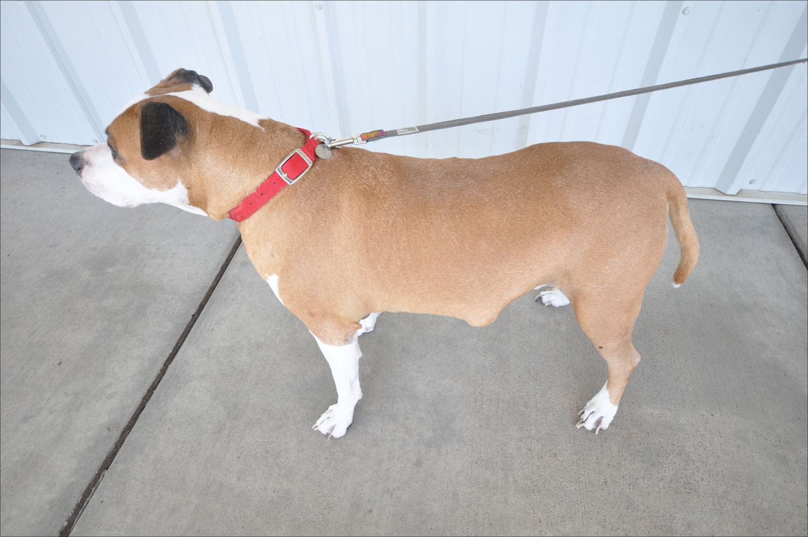
Given the opportunity to examine an older dog, I’ll very likely find at least one or two cutaneous (within the skin) or subcutaneous (just beneath the skin surface) lumps and bumps. Such growths are common by-products of the canine aging process. In this regard, I liken them to the brown spots that appear on our skin as we get older.
The good news is that most cutaneous and subcutaneous canine tumors are benign. It’s the small population of malignant masses that keeps us on our toes. They are the reason it’s important to have your veterinarian inspect any newly discovered lumps and bumps your dog develops. The smaller a cancerous growth is at the time of treatment, in general, the better the outcome.
Pet Your Dog to Find Tumors
In terms of “lump and bump patrol,” your first order of business is to pet your dog. No doubt you and your best buddy already enjoy some doggie massage time. What I’m asking you to do is a more methodical petting session. Once a month, slowly and mindfully slide your fingers, palm sides down, along your dog’s body. Move systematically from stem to stern while inspecting for any new lumps or bumps.
Also, look and feel for changes in the size or appearance of those previously discovered. Any new findings should be addressed with your veterinarian, who relies upon your help with this surveillance. Imagine your vet trying to find a tiny growth on a shaggy Sheepdog or Sheltie during the course of a single exam. Some lumps and bumps are bound to be missed without your assistance.
When to See Your Veterinarian
Does finding a new growth suggest that you must see your veterinarian right away? Not necessarily. Say that you’ve just spotted a new bump in your dog’s skin that is no larger than the size of a pea. She is due for her annual physical examination in three months. Must you rush to visit your vet with this new finding, or can it wait the three months? The answer depends on the behavior of this newly discovered growth.
My recommendation is that you continue to observe it once a week. Examining it more frequently can make it difficult to accurately assess change. If the mass is growing, or otherwise changing in appearance, best to have it checked out sooner rather than later. If no changes are observed, waiting to address it at the time of the annual physical exam makes perfectly good sense.
In contrast, say that in the course of examining your best buddy you discover a prune sized, firm, subcutaneous growth that feels attached to the shoulder blade. Based on the larger size and deep attachment of this mass, it’s best to have this one checked out right away. If ever in doubt, give your veterinarian a call to figure out the best course of action. As with most things medical, better to be safe than sorry.
In advance of your veterinary visit, mark the location of any lumps or bumps requiring inspection. You can clip some hair over the site or mark the fur with a ribbon, hair band, or marking pen. Growths discovered at home when an animal is lying down in a relaxed, comfortable position have a habit of magically disappearing when the dog is upright and uptight in the exam room.
Fine Needle Aspirate for Cytology
If a newly discovered growth is large enough, the usual first step your veterinarian will recommend is a fine needle aspirate for cytology. Cytology refers to the microscopic examination of cells. The purpose of this step is to attempt to noninvasively clarify the cell type within the mass, and whether it is benign or malignant.
Collection of a fine needle aspirate is a simple process that is easy on the dog and rarely requires any sort of sedation. Using a needle no larger than the size of a vaccination needle along with some gentle suction, your vet will remove a smattering of cells from the growth. These cells are then spit out onto a glass slide and evaluated under the microscope.
Some cytology interpretations are a slam-dunk, and can readily be interpreted by your family vet. Others require the eyeballs of a specialist – a clinical pathologist who works in a veterinary diagnostic laboratory. Remember, the goal of the cytology testing is to determine the underlying cell type, therefore whether the growth can be left alone or requires more attention. Fine needle aspirate cytology is often (but not always) definitive. If the results do not provide clarity, a surgical biopsy of the mass may be recommended.
If your veterinarian recommends surgical removal of a mass as the very first step (chooses to forego the fine needle aspirate), I encourage you to consider getting a second opinion. It is disappointing and frustrating when a veterinarian foregoes cytology, proceeds with surgery, and the biopsy report reveals a malignancy with cancer cells extending beyond the margins of the tissue that was removed. In other words, cancer cells were clearly left behind. Had the veterinarian known in advance from the cytology report that the tumor was malignant, a different approach (much more aggressive surgery and/or radiation therapy) would have been undertaken, almost certainly resulting in a better outcome.
A second “bad news scenario” that can arise from forging ahead with surgery without benefit of fine needle aspirate cytology is failure to identify a cancerous growth that may have already spread elsewhere in the body. If the cytology reveals a malignancy, screening the rest of the body for metastasis (spread) is a logical next step. If metastasis is discovered, removal of the originally discovered mass is unlikely to provide any benefit. Rather, such surgery will only subject the patient (and the client’s pocketbook) to a needless procedure. Leaping into surgery to remove a mass without the benefit of cytology is risky business.
Lipomas (Fatty Tumors)
Lipomas are one of the most common types of canine tumors. Fortunately, the vast majority of them are completely benign. They arise from fat (lipid) cells and grow in subcutaneous locations, primarily in the axillary regions (armpits) and alongside the chest and abdomen. Occasionally one will develop within the chest or abdominal cavity. Rarely does a dog develop only one lipoma. They tend to grow in multiples, and I’ve examined individual dogs with literally hundreds of lipomas.
Should lipomas be treated in some fashion? In the vast majority of cases, the answer is a definite, “No!” This is based on their benign, slow-growing nature. The only issue most create is purely cosmetic, which dogs could care less about!
There are a few exceptions to the general recommendation to let sleeping lipomas lie. A fatty tumor is deserving of more attention in the following situations:
A lipoma that is steadily growing in an area where it could ultimately interfere with mobility. The armpit is the classic spot where this happens. The emphasis here is on the phrase “steadily growing.” Even in one of these critical areas there is no reason to remove a lipoma that remains quiescent with no discernible growth.
Sudden growth and/or change in appearance of a fatty tumor (or any mass for that matter) should prompt reassessment to determine the best course of action.
Every once in a great while, a fatty tumor turns out to be an infiltrative liposarcoma rather than a lipoma. These are the malignant black sheep in the family of fatty tumors. Your veterinarian will be suspicious of an infiltrative liposarcoma if the fine needle aspirate cytology reveals fat cells, yet the tumor feels fixed to underlying tissues. (Lipomas are normally freely moveable.) Liposarcomas should be aggressively surgically removed and/or treated with radiation therapy.
Occasionally a lipoma grows to truly mammoth proportions. If ever you’ve looked at a dog and thought, “Wow, there’s a dog attached to that tumor!” chances are you were looking at a lipoma. Such massive tumors have the potential to cause the dog discomfort. They can also outgrow their blood supply, resulting in potential infection and drainage from the mass. The key is to catch on to the mass’s rapid growth so as to surgically remove it before it becomes enormous in size and far more difficult to remove.
How can one prevent canine lipomas from occurring? No one knows. Anecdotally speaking it is thought that overweight dogs are more predisposed to developing fatty tumors. While I’m not so sure I buy this, I’m certainly in favor of keeping your dog at a healthy body weight.
Sebaceous Adenomas
These are the most common benign skin tumors in dogs. Sebaceous glands are microscopic structures found just beneath the skin surface. They secrete an oily substance called sebum that is transported to the skin surface via microscopic ducts, and can arise from the gland or the duct.
Sebaceous adenomas tend to be small, no more than 1/4 to 1/3 of an inch in size. They may appear round, or they can have a wart-like appearance. Sebaceous adenomas occur primarily in middle-aged and older dogs. Any breed can develop sebaceous adenomas, but certain breeds are particularly predisposed: English Cocker Spaniels, Cocker Spaniels, Samoyeds, Siberian Huskies, Alaskan Malamutes, West Highland White Terriers, Cairn Terriers, Dachshunds, Miniature Poodles, Toy Poodles, Shih Tzus, Basset Hounds, Beagles, and Kerry Blue Terriers.
Because of their benign nature, the vast majority of sebaceous adenomas require no treatment whatsoever. There are some exceptions to this general rule, and they are as follows:
Surgical removal is warranted for those sebaceous adenomas that recurrently bleed or become infected because of self-trauma (the dog bites or chews at them) or because they get in the way of the groomer’s clippers.
Some sebaceous adenomas secrete oodles of sebum, creating the constant appearance of an oil slick on the dog’s hair coat. The grease rubs off on hands, furniture, and anything else the dog contacts. No fun!
Some sebaceous adenomas are pretty darned unsightly, looking like warty little aliens poking through the hair coat. Although this is not bothersome for the dog, it can be a significant issue for the person living with that dog.
If a mass believed to be a sebaceous adenoma is growing or changing in appearance, it is important to ask your vet to have another look. What was thought to be a benign adenoma may be its less common cancerous cousin, a sebaceous carcinoma.
The Importance of Histopathology for Your Dog
If your veterinarian surgically removes a growth from your dog, do not, I repeat, do not let that tissue sample wind up in the vet clinic garbage can! A far better choice is to have the mass submitted for histopathology (biopsy) to a veterinary diagnostic laboratory. There, a veterinary pathologist will evaluate paper-thin slices of the mass under the microscope to confirm the identity of the mass.
Even if fine needle aspirate cytology indicated that the growth was benign, histopathology is warranted. On occasion, the pathologist discovers something quirky such as a malignant tumor within the center of a benign lipoma.
If histopathology is not affordable, ask your vet to place the growth that was removed in a small container of formalin (preservative) that you can take home for safekeeping. This way, should multiple masses begin growing at the surgery site or should your dog develop a tumor at another site, you will still be able to request histopathology on the original sample. Formalin is toxic stuff, so keep the container lid sealed tightly.
Lumps and bumps are a very normal part of the canine aging process. Teaming up with your veterinarian to assess them on a regular basis is the best way to insure that they never create a health issue for your best buddy.