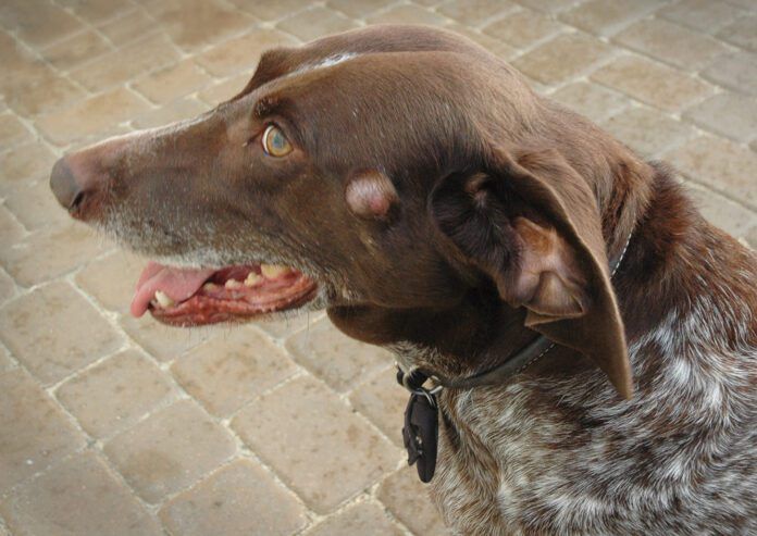
Finding a skin lesion or lump that was not there the last time you caressed your dog can instantly transform your mood from contented to fearful. Is it cancer? Many skin lesions and lumps look cancerous.
If it’s any comfort, you should know that few skin lesions turn out to be cancer – but that said, anything that is new or that has recently changed should be brought to the attention of your dog’s veterinarian, who can determine what’s up with the lump.
The veterinarian will examine the skin lesion, check your dog for any other lesions, and obtain a history from you about other relevant aspects of your dog’s health.
Although some skin lesions have a characteristic appearance that helps a veterinarian identify them as the most likely type of lesion or tumor, a definitive diagnosis can be made only by fine needle aspirate (FNA) with cytology (examining some of the cells that were removed with the needle under a microscope) or biopsy with histopathology (removing some tissue from the lesion and examining it under a microscope).
(See sidebar “Cytology and Biopsy,” for more information about how samples of your dog’s lesion may be collected and examined in pursuit of a diagnosis.)
Common Canine skin lesions
As you read this section about the most common skin lesions found on dogs, keep in mind that they may sound lethal, but that’s not necessarily so. Learning about them and understanding the best course of action for each will help lessen your fears. This information is designed to help you discuss the lesions with your veterinarian, so the two of you can determine what’s best for your dog.
- Benign lipomas
Often referred to as “fatty tumors,” benign lipomas are solitary deposits of fat underneath the skin. They are round, smooth, and a little squishy. These are not cancerous tumors and do not need to be removed unless they are impairing a dog’s ability to move.
Benign lipomas can grow anywhere on the body, but are predominantly found on the chest, belly, and upper limbs. They begin as small masses, often the size of a marble or golf ball. Some lipomas change very little in size over a dog’s lifetime but some can grow to be the size of a cantaloupe.
- Mast cell tumors
Mast cells are white blood cells; they are found in many areas of the body but are particularly prevalent in the skin. They are one of the cells involved in allergic responses and release histamine when activated.
A mast cell tumor is a proliferation of mast cells that forms a nodule on the skin. These tumors can be flat or raised; flesh-colored, pink, or red; and may be ulcerated. They may grow rapidly in as little as a week or may remain unchanged in size for months.
If your veterinarian suspects that your dog’s lesion is a mast cell tumor, an injection of diphenhydramine (Benadryl) may be given before aspirating it. When a mast cell tumor is poked or disturbed, it may release large amounts of histamine. This can cause anaphylaxis (a life-threatening allergic reaction)or can lead to the development of stomach ulcers. Diphenhydramine minimizes the risk of these events. Your veterinarian also may instruct you on how much Benadryl to give at home until the cytology results have been returned.
Mast cell tumors are often successfully diagnosed by FNA and cytology. But determining if the mast cell tumor is low grade or high grade requires biopsy and histopathology. Your vet will likely recommend surgical excision of the lesion if it is determined to be a mast cell tumor.
Mast cell tumors tend to have longer, microscopic roots traveling underneath the skin than other types of skin lesions. Your veterinarian may recommend that your dog see a board-certified veterinary surgeon for removal of the mass so that wider surgical margins can be obtained.
Mast cell tumors are more likely to be a low grade tumor than a high grade tumor. Low grade tumors are typically cured with complete surgical excision. High grade tumors are more likely to recur, either in the same location or in different locations. Dogs that have a high grade mast cell tumor would benefit from a consultation with an oncologist and may require chemotherapy with or without radiation therapy.
Although we do not know what causes mast cell tumors to grow, there appears to be some genetic mutations that increase the risk for developing mast cell tumors. Certain breeds of dogs – like the Boxer, Boston Terrier, and Labrador Retriever – are at increased risk for mast cell tumors.
Mast cell tumors can also affect the nail bed and internal organs, such as the spleen.
- Cutaneous histiocytomas
Like mast cells, a cutaneous histiocytoma is a proliferation of cells involved with the immune system. Instead of mast cells, however, histiocytomas are composed of a histiocyte called a Langerhans cell.
This is a benign tumor that often has a similar appearance to mast cell tumors. Histiocytomas are most commonly found in dogs less than 6 years old. They initially grow rapidly and then often remain the same size until they spontaneously regress and disappear a few months later.
Histiocytomas are often readily diagnosed by FNA and cytology. Because they can appear similar to mast cell tumors, it is important to complete this diagnostic test to confirm the type of lesion. If the lesion is a histiocytoma, no further intervention is necessary. It will regress and resolve on its own.
- Squamous cell carcinomas
Squamous cell carcinomas are tumors of the squamous cells in the epidermis, or top layer of skin. This tumor can be a small, raised area on the skin surface that looks like a wart but is often red, irritated, or ulcerated.
Squamous cell carcinoma is most often found on areas of the body that have a thinner hair coat, like the ears, near the lips or along the top of the nose, and the underside of the belly. Dogs with short-haired coats, and particularly those with light-colored coats, are at increased risk for developing squamous cell carcinoma.
Although the direct cause of squamous cell carcinoma is unknown, it is suspected that exposure to sunlight and other forms of ultraviolet (UV) light increases the risk of developing squamous cell carcinoma.
Squamous cell carcinoma often appears as a single lesion on the skin. Surgical removal of the tumor is the treatment of choice as these tumors tend to be locally invasive and don’t typically spread.
There are two forms of squamous cell carcinoma that are more serious than the singular lesions that can appear on a dog’s skin. The first is multicentric squamous cell carcinoma. This is a very rare condition of dogs in which more than one lesion appears on multiple areas of the body.
The second is squamous cell carcinoma of the nail bed. This appears first as a swollen digit and can cause deformation or loss of the claw. This form of squamous cell carcinoma is metastatic, meaning that it can spread to other areas of the body, such as the lungs. Presurgical staging and amputation of the affected digit is often recommended.
- Melanomas
Melanoma is a type of skin cancer that is derived from the abnormal growth of melanocytes, the pigmented cells of the skin. Although we don’t know the cause of melanomas in dogs, there is an association between high levels of UV exposure and the development of melanomas in humans.
There are three different forms of melanoma in dogs. Cutaneous melanoma is typically a brown or black lesion that has a flat or wrinkled appearance. There is a form of this melanoma called amelanotic melanoma that contains no pigment and may be flesh-colored. Cutaneous melanoma tends to be benign in dogs and surgical excision of the skin lesion is typically curative.
The other two types of melanoma in dogs are oral melanoma and digit melanoma. These are typically malignant tumors that can spread to other areas of the body. Treatment includes extensive surgical excision of the mass; for an affected digit, this includes amputation. Histopathology of the tumor can determine the grade of melanoma and appropriate follow-up care with an oncologist. A melanoma vaccine called ONCEPT may be recommended to train your dog’s immune system to find and eliminate malignant melanoma cells.
Melanoma tumors do not exfoliate well when aspirated. If your veterinarian suspects that a lesion may be a melanoma, then a surgical incisional or excisional biopsy may be recommended instead of an FNA.
- Glandular and follicular tumors
These are tumors that arise from the glands within the skin or from within the hair follicle. They look like a fleshy, raised irregular growth and are often called warts because of their appearance.
These tumors can grow and shrink in size over time. When the tumor grows, it often fills with a thick caseous fluid that resembles cottage cheese. The tumor will often burst, or erupt, releasing this thick fluid. As the tumor dries out, it shrinks but never completely disappears.
Glandular and follicular tumors are typically benign but can be malignant in rare cases. An FNA of the mass will often confirm that it is a glandular or follicular tumor but is usually not good at determining if it is malignant or benign. Surgical excision with histopathology of the mass is often needed to confirm the type and determine if follow-up care with an oncologist is necessary.
- Cutaneous lymphoma
Lymphoma is a cancer of lymphocytes, which are a type of white blood cell. When we think of lymphoma in dogs, we often think of the condition that causes enlarged, painful lymph nodes or that creates tumors on the spleen, liver, or other organs in the abdomen or chest. However, lymphoma can appear anywhere there are lymphocytes; this includes the skin.
Cutaneous lymphoma appears as small red patches on the skin that are often circular or oval shape. These patches may look irritated or ulcerated. They can look similar to skin lesions caused by other diseases, like atopic dermatitis. The cause of cutaneous lymphoma is unknown.
Dogs with cutaneous lymphoma may have decreased energy and appetite. Cutaneous lymphoma can produce a protein that increases the calcium level in a dog’s blood. The increased calcium causes the dog to drink more water and urinate more.
Cutaneous lymphoma is diagnosed by incisional biopsy of one or more skin lesions. The treatment of choice is chemotherapy.
- Fibrosarcomas
A fibrosarcoma is a tumor of the connective tissue in the body. Connective tissue is found in many areas of the body, including the skin.
A fibrosarcoma may appear as a firm nodule within or just underneath the skin. The skin over the mass will likely appear normal, but the mass will feel like a firm nodule the size of a pea or a marble.
Although the cause of fibrosarcomas is not known, it is suspected that previous trauma is a potential risk factor. Previous trauma may include an injury to the skin, foreign material (like a grass awn or foxtail) within or just underneath the skin, or any type of injection (this is more common in cats than dogs).
Most fibrosarcomas in dogs are benign and slow to grow. However, even benign fibrosarcomas tend to be locally invasive. It is best to surgically remove this type of tumor when it is small and can be more easily removed. Pre-surgical staging may be necessary to determine the extent of the tumor. Referral to a board-certified veterinary surgeon may be recommended to achieve the best possible outcome.
Don’t panic!
Finding a new skin lesion on your dog can be concerning. Thankfully, most skin lesions are benign but should be addressed in a timely manner. Your veterinarian will guide you through the process of diagnosing and treating any new skin lesion you find.
Fine needle aspiration (FNA) involves inserting a needle into the lesion to obtain a sample of cells. The sample is sprayed onto a slide and submitted to a laboratory for a veterinary pathologist to read. The needle used to obtain the sample is no bigger than the one used to give your dog a vaccine. Your dog will feel only a small pinch during the FNA procedure.
The cytology will return one of three results: a definitive answer as to the type of lesion; the class of lesion but no information about whether it is benign or malignant; or no definitive answer at all. Some lesions do not exfoliate well – they don’t release any intact cells to examine. Other lesions bleed easily when they are aspirated. When this happens, there is so much blood present on the slide that cells from the lesion cannot readily be seen under a microscope. This is called hemodilution.
Surgical biopsy (also called an incisional biopsy) is where a sample of the skin lesion is surgically removed. An incisional biopsy may be recommended if the lesion is in a location where removing all of it may be difficult or if there are multiple lesions that appear identical. The sample is sent out to the laboratory for histopathology (examination under a microscope after they have been stained with dye).
Surgical excision (also called an excisional biopsy) is where the entire skin lesion is removed and sent out for histopathology. Your veterinarian will need to make an elliptical incision around the lesion that is at least 1 centimeter wider on either side to remove all of the lesion. Skin lesions tend to extend their roots underneath the skin’s surface, much like a tree extends its roots below the ground. An elliptical incision is easier to close with a better cosmetic outcome than a circular incision.
Histopathology of the lesion helps to determine what additional treatments may be necessary. For an incisional biopsy sample, histopathology determines if the lesion needs to completely removed and if any presurgical imaging is required, such as radiographs, ultrasound, or CT scan. For an excisional biopsy, histopathology determines if clean margins were obtained when removing the lesion. It also helps guide any need for oncology care or further monitoring following removal of the lesion.


