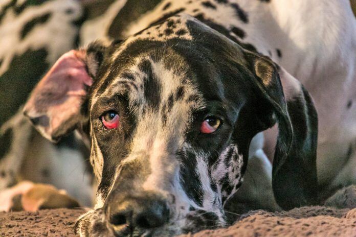
Horner’s syndrome is a neurologic condition that affects your dog’s eye and face, usually on just one side. It helps to have a “normal” side to compare the changed side to. You notice changes when you look at your dog’s face.
It’s not the same as a cross-eyed dog or cherry eye, although cherry eye has some similar features.
With Horner’s syndrome:
- The upper eyelid will droop down
- The eye may seem sunken
- The pupil of the affected eye will be smaller than the pupil in the other eye
- The dog’s third eyelid will be up (partially blocking the eye)
- The eyelid may be red
What Causes Horner’s Syndrome in Dogs?
The cause of Horner’s is normally an injury or irritation to the nerves around the eye, but often the exact insult is not determined. Obvious causes would be a bite wound or a tumor affecting those nerve roots.
Internal ear infections can also irritate the nerves running to those areas. The nerves involved cover a wide range of your dog’s body: chest, head, and neck. That means lots of places for the nerves to be affected.
Because possible causes of Horner’s syndrome include intervertebral disc disease, cervical myelopathy, an inner ear infection, or any trauma, it’s wise to contact your veterinarian.
Sometimes, the cause is unknown (idiopathic). A 2000 study published in the Journal of Neuro-Ophthalmology looked at 155 dogs, 110 of which were Golden Retrievers, with Horner’s syndrome. They found 100 of the Goldens were suffering from idiopathic Horner’s.
What to Do About Horner’s in Dogs
There is no specific treatment, other than time, for Horner’s. Many cases resolve on their own over time, although some stay. Horner’s is not painful. Most dogs function just fine despite their slightly “loopy” appearance.
The Dog’s Third Eyelid
One side effect of Horner’s is that you will now notice your dog’s third eyelid. Normally, the third eyelid is pale and barely noticeable. Some will be pigmented, but they still fit in with the lower lid.
The third eyelid, or nictitating membrane, can have some problems of its own, such as “cherry eye.” When you look at your dog you see a pink or red swelling on the inner corner of your dog’s eye. In cherry eye, the third eyelid and its gland that helps to lubricate the eye, prolapses.
Cherry eye is often bilateral and more common in brachycephalic breeds with short faces. This is usually evident in young dogs. Massage and eye ointments or drops to minimize inflammation may work to stop this condition. More commonly, it will recur, especially in times of allergies such as pollen season.
Surgical Correction for Horner’s Third Eyelid
Your veterinarian can tighten up the tissues surgically to help keep the third eyelid in place. Your veterinarian may refer you to a veterinary ophthalmologist for that.
While the appearance of a prolapsed third eyelid may be disturbing, the condition is not serious or painful. It does warrant a veterinary visit but not an emergency clinic run.


