


Dogs with hip dysplasia can lead a relatively normal life, but early intervention is the key to maintaining pain-free mobility and an active lifestyle. Even dogs with arthritis secondary to hip dysplasia can have an improved quality of life with surgical intervention or medical management.
What is hip dysplasia in dogs?
Discovered in 1935, hip dysplasia is one of the most common genetically inherited orthopedic traits. Fortunately, breeders are aware of this fact, and reputable breeders avoid breeding dogs with this defect. However, it is not always possible to know for certain when choosing breeding dogs.
Hip dysplasia is the malformation of the hip joint during a puppy’s growth period. The hip joint is composed of two parts: the head of the femur (ball) and the acetabulum (socket). When a puppy grows, the head of the femur and the acetabulum normally grow at a uniform rate. This creates a snug fit of the ball (femur) within the socket (acetabulum).
In hip dysplasia, the head of the femur and the acetabulum grow at different rates, which creates laxity in the joint. That loose fit results in abnormal movement, which eventually creates scar tissue along the edges of the cartilage. A hip joint that is lined with scar tissue is an arthritic joint.
Signs of Hip Dysplasia in Dogs
Some puppies who have hip dysplasia may walk with a bunny hop gait in their hind limbs. While limping is an obvious sign of dysplasia, if both rear legs are affected, you may not notice it. While many puppies with hip dysplasia show no overt signs of lameness or gait abnormalities, you may notice one puppy is less active or even seemingly clumsier than his littermates. He may walk wobbly, have uneven muscle development in the rear end, or walk with a slight rear-end “sway.”
Puppies with dysplasia often are less active due to soreness. An affected pup may have more trouble doing stairs than littermates. While most puppies don’t show acute pain, some will. Those pups may snap or cry if you touch or try to manipulate the painful hip joint. Astute owners may notice a difference in muscle mass between the two rear legs if only one hip is involved.
As dogs with hip dysplasia develop arthritic hip joints, they may show periodic lameness, a reluctance to use stairs, and may be slow to rise. They will often have a shortened stride in their hind limbs or have atrophied thigh muscles because they cannot fully extend their hips. Of course, not all arthritic hip joints are caused by hip dysplasia, and signs of hip dysplasia can be like those caused by osteoarthritis in joints other than the hips.
Does My Puppy Have Hip Dysplasia?
A simple veterinary palpation technique can screen for hip dysplasia that can be completed during a puppy’s wellness exam. Called the Ortolani Sign, this palpation checks for laxity of the hip joint. Light sedation may be needed to relax the muscles around the hip (and to better control a wiggly puppy!).
A positive Ortolani Sign indicates the presence of hip dysplasia. However, some puppies with hip dysplasia may have a negative Ortolani Sign. An X-ray is needed if there is suspicion of hip dysplasia.
The gold standard X-ray for diagnosing hip dysplasia is an extended ventrodorsal view of the pelvis. It’s a single picture of your dog’s hips while he is lying on his back with his hind limbs extended. This radiograph can be interpreted by a board-certified veterinary radiologist or submitted to the Orthopedic Foundation for Animals (OFA) for analysis and certification.
Unfortunately, this method does not consider the forces that apply pressure to the hips when a dog is standing. Therefore, the hips of some dogs with hip dysplasia may appear normal using this method. Also, this view may not be diagnostic in dogs who are less than 1 year old.
In contrast, the University of Pennsylvania Hip Improvement Program (PennHIP) method can be used to detect if a puppy is likely to develop hip dysplasia and can be used in puppies as young as 4 months old.
The PennHIP method takes three views of your dog’s hips. The first view is the extended ventrodorsal view discussed above and is used to detect if any arthritic changes are evident in the hip joints. The second view simulates how the hip joints would appear if your dog were held up in a standing but non-weight position. The third view simulates how your dog’s hip joints appear if he were standing and bearing weight on his hind limbs.
What Causes Hip Dysplasia in Dogs?
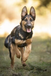
Any breed of dog can have hip dysplasia, although it is more commonly seen in large-breed dogs such as German Shepherds, Labrador Retrievers, Golden Retrievers, and Saint Bernards.

Hip dysplasia is a hereditary condition that is also influenced by:
- Nutrition: All puppies should be fed either a food developed for “all life stages” or one specified as a “puppy” food. Large breed puppies need a high-quality large-breed puppy food that specifically states it is formulated for large-breed puppies. A puppy food formulated specifically for large breeds delivers a more controlled amount of calcium and protein to ensure proper bone development.
- Exercise: Strenuous exercise in puppies, such as long hikes or going for long runs, can lead to premature excessive muscle development. This can contribute to laxity of the hip by changing the mechanical forces on the joint as it develops. Discuss appropriate exercise for your puppy at the puppy’s wellness exam. High-action games like fetch may not be recommended.
- Growth Rate: All puppies regardless of breed or size should be kept lean as they grow and not allowed to gain excess weight. Being overweight puts additional strain on the hip joints and promotes joint laxity.
Surgical Options for Hip Dysplasia in Dogs
Puppies who have been diagnosed with hip laxity between 10 and 18 weeks of age may benefit from a surgical procedure called Juvenile Pubic Symphysiodesis (JPS). This procedure prematurely closes a growth plate in the bottom of the pelvis. Closing this growth plate causes the acetabulum to cup the head of the femur more as it grows over the next four to six months, minimizing joint laxity and the development of hip dysplasia. Puppies with hip laxity who are older than 18 weeks of age are not candidates for this procedure.
Puppies between 4½ and 10 months of age who have been diagnosed with hip dysplasia may benefit from a procedure called a double or triple pelvic osteotomy (DPO or TPO). In this surgical procedure, two or three cuts are made in the pelvis. Then the acetabulum is rotated so that it properly cups the head of the femur.
Only puppies with mild to moderate hip laxity qualify for the DPO or TPO procedure. Puppies with severe hip laxity, lameness, or evidence of arthritic changes to their hips on radiographs should not undergo a DPO or TPO.
Adult dogs with hip dysplasia have two surgical options to improve their quality of life and give them pain-free movement of their hips. These procedures are total hip replacement (THR) or femoral head osteotomy (FHO).


I have OFA’d my dogs since the early 90s and they have never been under any type of anesthesia. Any vet who requires it, find a different vet. There are plenty who will do it without. PennHip, yes, a different matter. Anesthesia, plus the grading is complicated if there aren’t enough dogs in the data base to compare to. Advantage with PennHip is that it can be done at a much younger age rather than wait till age two with OFA for certification.