


Mast cell tumors (MCTs) are the most common form of malignant skin cancer that occurs in dogs, accounting for about 14 to 21 percent of all skin tumors. There is significant variability in the presentation of MCTs from dog to dog and even from tumor to tumor, which explains why this form of canine neoplasia is often referred to as complicated and challenging. But the fact remains that MCTs are treatable, and many dogs are considered cured after treatment and go on with happy, healthy lives.
How Mast Cell Tumors in Dogs Develop
Mast cells are a subtype of white blood cells that derive from the bone marrow and then become residents of specific tissues, especially those that are closest to the external surfaces of the body, such as the tissues of the nose, mouth, lungs, skin, and around blood vessels. Their primary functions include defense against parasitic infection, tissue repair, and formation of new blood vessels; they also play an important role in the allergic response system. They can be activated by the immune system – a process called degranulation – to release a wide variety of inflammatory mediators, including histamine and heparin, which in turn modify immune reactions and inflammation.
When mast cells replicate in higher than normal numbers, an MCT (also known as a mastocytoma) can form. They are usually first noticed on or just under the skin, but they can form anywhere on the body. The most common location is on the trunk, followed by the limbs and in between the toes; they can occasionally be found on the head and neck. The skin mass might appear as a raised lump or bump or growth; it can soft or firm; hairless or covered by hair; it may or may not be red, ulcerated, or swollen.
MCTs can also occur internally, developing on the spleen, liver, intestine, and in bone marrow, as well as on the salivary gland, nasopharynx, larynx, oral cavity, gastrointestinal tract, ureter, and spine. This type is more aggressive, and it is usually preceded by dermal and subdermal tumors.
MCTs can not only vary in location, appearance, shape, and size, but also have a tendency to present with varying clinical behavior, which is suspected to be influenced by breed. MCTs can even change in size (increasing and decreasing from day to day!) because of reactions occuring in the mass. This is due to secondary inflammation caused by the release of histamine, which then results in intermittent swelling.
This type of tumor tends to present in dogs of middle age or older, but can occur in dogs of any age. No gender predilection has been reported. MCTs typically present as a solitary tumor but about 11 to 22 percent present with more than one tumor and 10 to 20 percent of those that have had an MCT will develop a new MCT in another location at some point.
The Cause of Mast Cell Tumors
The cause of MCTs is unknown. Certain breeds of dogs are predisposed to them, so there may be some genetic component. A genetic mutation in the protein c-kit tyrosine kinase receptor – called a c-kit oncogene – is found in 25 to 30 percent of tumors. Chronic inflammation may also predispose dogs to developing the disease, such as dogs that have a history of allergic skin disease. There has been no association found between sun exposure and MCT in dogs.
Breeds With a Predisposition for Mast Cell Tumors
MCTs are over-represented in Pugs (at risk for multiple but low-grade tumors), Boxers, Labrador Retrievers (at increased risk for the more aggressive form), Golden Retrievers, Beagles, and Boston Terriers. Other breeds showing an increased likelihood of developing the disease include Schnauzers, Bullmastiffs/Bulldogs/ Bull Terriers, Cocker Spaniels, Fox Terriers, Weimaraners, Rhodesian Ridgebacks, Shar pei, and Dachshunds.
Diagnosis of Mast Cell Tumors
The initial evaluation of a suspected MCT begins with a fine needle aspirate and cytology, from which a diagnosis is usually made. Grading of the tumor cannot be done at this point as only cells are obtained from the aspirate; visual examination combined with the cytology report will determine if the MCT is a candidate for surgical excision.
It is reasonable to stage (evaluate how far the disease has spread) the tumor prior to surgery; in cases of a recurring tumor, multiple tumors, or evidence of metastasis, staging in advance is considered necessary.
When an MCT spreads, the cancer usually moves into the regional lymph nodes near the tumor, then to the liver and spleen; it can also spread to other places in the abdomen as well as to the bone marrow. At a minimum, the staging tests should include an aspirate of the lymph nodes near the tumor and an abdominal ultrasound.
The ultrasound can show if there is any evidence of metastasis or anything suspicious that requires further testing. Aspirates of the liver and spleen may be recommended, even if all the organs appear normal, because they may harbor cancer cells.
Treatment for Mast Cell Tumors in Dogs
The preferred treatment for MCTs is surgical removal – and for many cases, this is the only treatment needed. Prior to surgery and to alleviate some of the unpleasant symptoms of MCTs, a dog may be placed on antihistamines and antacids to mediate the effects of the histamine generated by the mast cells. Because MCTs are prone to growing into surrounding tissues, the visible tumor is removed with the addition of wide surgical margins – 2 to 3 centimeters on all sides, as well as layer of tissue below – in an attempt to remove all cancerous cells; as a result, the surgical sites can be quite large. If there is more than one tumor, a biopsy is recommended for each one because individual tumors can be of different grades and require different courses of treatment.
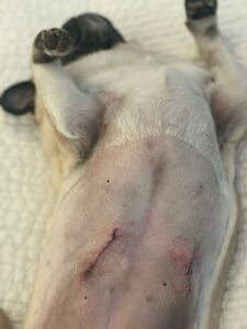
The excised tumor(s) will be sent to the lab for pathology; the resulting biopsy report will provide the tumor’s grade, confirm whether or not the tumor was completely removed (“clean” or “dirty margins”), and provide a foundation for prognosis. If the biopsy report shows that detectable cancer cells remain, the tumor can regrow. In these cases, a second surgery (if the site is amenable) or radiation therapy can be recommended. Mast cell tumors can be slower to heal post-operatively with suture sites that have the potential to break down.
If the location of the lesion disqualifies it as a candidate for surgery, further diagnostics can be performed to obtain its disease stage. If the tumor is deemed too large for removal, chemotherapy and/or radiation treatments may be recommended in an attempt to reduce the mass to an operable size. Inoperable tumors should be evaluated by incisional biopsy for histologic grade.
The decision to pursue additional treatment should consider the grade of the cancer as well as the results of other prognostic tests. Post-surgical radiation and chemotherapy are warranted on a case-by-case basis. The most important considerations when determining if a patient needs additional treatment following surgery are tumor grade, how complete the excisions of the tumor were, and whether the MCT has spread or not.
Mast Cell Tumor Grading
Traditionally MCTs have been classified using the Patnaik grading system, which assigns a histological grade of I, II, or III to each tumor. Studies have shown that the grade assigned to a particular tumor can vary as it is based on the subjective opinion of the pathologist; accordingly, sometimes a second evaluation of the biopsied tissue is warranted.
- Grade I. About 33 to 50 percent of MCTs are classified as Grade I; these tumors act in a benign manner and are usually considered cured with complete surgical excision and simple monitoring over time. They tend to be contained and not prone to metastasis or recurrence. One exception is low-grade tumors on the dog’s muzzle or lips; these have a high rate of spread and need to be treated more aggressively, as 50 to 65 percent of them will develop metastasis.
- Grade II. About 25 to 45 percent of MCTs are classified as Grade II – and this is where the decisions about their treatment get really challenging. Some Grade II tumors act like Grade I tumors, but others are similar to Grade III and more difficult to treat.In general, Grade II tumors are less defined than Grade I tumors and about half of them are more likely to spread into surrounding tissues and other parts of the body, with about 25 percent having already spread at the time of diagnosis. A Grade II tumor with a mitotic index (more on this below) of greater than 5 should be treated as a Grade III tumor.
- Grade III. About 20 to 40 percent of MCTs are classified as Grade III. These very aggressive tumors invade quickly and into deep tissue layers. About 50 to 90 percent grow and metastasize, with most cases showing spread by the time of diagnosis. If removed, 55 to 95 percent are likely to recur. Surgery along with chemotherapy and/or radiation is usually recommended.The Kiupel system of MCT grading is more recent and achieves better consensus among veterinary pathologists. This simpler approach categorizes the tumors as either high-grade or low-grade; the assessments this system uses provide for a more accurate prediction of the behavior of the disease and therefore hopefully to determine a more significant prognosis. High-grade tumors tend to be associated with increased likelihood to metastasis and poor prognosis with an average survival time of approximately four months. Low-grade tumors have a median survival time of more than two years.
Supplemental Testing
As there is no single factor or test that accurately predicts the behavior of the disease, supplemental testing can be invaluable in developing an accurate assessment and improving the prognosis of affected dogs.
- Mitotic Index. This test measures the rate at which the malignant mast cells are dividing and populating at the time of biopsy (this is one of the tests in the Kiupel system of grading) and is now usually included in biopsy reports. A tumor with a mitotic index of 5 or less can be treated as a Grade I MCT with a good prognosis and a median survival time of more than five years (regardless of histological grade). MCTs with a mitotic index over 5 should be treated as Grade III tumors. Median survival time for those with higher mitotic indexes is two to four months, again, regardless of the grade.In general, the higher the mitotic index, the poorer the prognosis. For those tricky Grade II tumors especially, this test provides insight into how the tumor is likely to behave, so an appropriate treatment plan can be developed.
- MCT Panel. The MCT prognostic (or proliferation panel of tests can derive additional information from the biopsy sample may help develop a prognosis and treatment plan, especially for high-grade/Grade II/Grade III tumors. This panel evaluates the specific markers AgNOR, PCNA, Ki-67, c-Kit, and tests for c-Kit mutation status, which are related to the proliferation of the MCT.
Approximately one-third of all dogs with MCTs have c-Kit mutation of the mast cells. This is an abnormality in the c-Kit gene, which affects a protein found on the cell’s surface and is involved with proliferation and other biological activities; the mutation causes the tyrosine kinase receptor enzyme in the gene to be stuck in the “on” position, causing uncontrolled growth. In particular, the c-Kit mutation test can indicate whether chemotherapy is warranted, as well as provide a guideline for which chemotherapy protocol to use.
While the presence of this mutation is usually found in higher-grade tumors and indicates a more aggressive form, it also may be more susceptible to treatment with a class of drugs known as KIT inhibitors. Palladia and Kinavet are two chemotherapy drugs that target this mutation specifically; they act by cutting of the blood supply to the tumor and inhibiting tyrosine kinase (a protein that plays a role in growth and development).
Other Treatments
MCTs are very susceptible to radiation therapy, a localized treatment. Radiation is especially helpful in preventing regrowth for incompletely excised tumors. The majority of dogs with low-grade MCT remain tumor-free two to five years after surgery and radiation therapy.
Even dogs with Grade III MCT can benefit from this dual mode treatment; one study documented 70 percent still alive one year following treatment; another study reported a median survival time of 20 months. It may also be used to treat tumors that are not candidates for surgical removal due to size and/or location, either to reduce the size prior to surgery or as palliative care by reducing the size of the tumor and improving clinical signs. It does not prevent metastasis.
Radiation as a primary treatment is considered a palliative approach; the addition of chemotherapy and steroids can improve this approach and may be offered for cases with no other treatment options.
Chemotherapy may be recommended as part of the treatment protocol in cases where the MCT has metastasized or has been rated as high-grade or Grade III, as well as those with a positive c-Kit mutation result, dirty margins, high scores on either the prognostic panel or mitotic index, or presenting with multiple tumors. Chemo may also be considered to prevent tumor recurrence, especially when radiation is not an option.
Combining chemo with surgery typically improves the prognosis of dogs with high grade tumors; median survival for dogs with Grade III tumors undergoing surgery alone is six months while those who undergo surgery and chemo have a median survival time of 12 months. Combination chemotherapy protocols, often in conjunction with steroids, also offer improved efficacy. Commonly used drugs include Lomustine (CCNU), Vinblastine, Palladia, and Kinavet. Vincristine, L-asparaginase, and cyclophosphamide have also been used with some effectiveness.
Supportive Treatment
In addition to antihistamines, supportive medications to consider include: prednisone to reduce inflammation; cimetidine, an antacid that helps to counter the increased acid production in the stomach caused by the MCTs; and intralesional triamcinolone, a corticosteroid, which can be injected directly into the tumor to reduce size. Cryotherapy is sometimes used to freeze and destroy small MCTs; this approach can be an option for older dogs for whom anesthesia and surgery are not an option.
Prognosis
Prognostic considerations for MCTs include grade, clinical stage, location, systemic symptoms, status of surgical margins, and mitotic index. Mast cell tumors that are completely removed and are Low-Grade, Grade I, or Grade II with a low mitotic index and free of metastasis have an excellent prognosis, with most cases being considered cured. Dogs with tumors that were incompletely removed but subsequently treated with radiation therapy have an excellent prognosis as well, with 90 to 95 percent having no recurrence of the tumor within three years.
Poor prognosis is associated with MCTs that occur on the muscle, around the mouth, in internal organs, in the bloodstream, or in bone marrow; those that are ulcerated, large, fast-growing, or recurring are also in this category. Dogs with c-Kit mutation are also associated with poor prognosis, as are dogs with Grade III tumors as local recurrence and/or spread is likely (only about 10 percent of these dogs live a year past surgery). Survival rates for dogs with a mitotic index of over 5 is only two to four months. As daunting as some of these figures are, remember that these are only guidelines; every dog is different.
On the Horizon
A number of exciting therapies for MCTs are in development:
- New treatments using receptor tyrosine kinases inhibitors for tumors with c-Kit mutations are an active part of cancer treatment research for both humans and dogs.
- QBiotics has completed clinical trials on Tigilanol Tiglate EBC-46, a novel anticancer pharmaceutical protein kinase C activator isolated from the seeds of the Blushwood tree (Fontainea picrosperma). The drug, administered by injection into the tumor, stimulates the immune system, resulting in destruction of the mass as well as the blood supply to the tumor. Complete tumor destruction was achieved in 75 percent of the cases after a single injection; the trial demonstrated that the treatment was well-tolerated (minor side effects) with rapid healing and minimal scarring. Application for approval is in process.
- A recent study at the Animal Health Trust Center for Small Animal Studies looked at the genetic changes in mast cells that promote metastasis. Researchers hope this will help develop a highly accurate test that can predict whether an MCT will metastasize; if successful, targets can be identified for new drugs to prevent metastasis. Future research will attempt to identify the causes of those genetic changes, resulting in the development of new cause-targeted anti-metastasis drugs.
- The Cummings School of Veterinary Medicine at Tufts University is conducting a clinical trial to evaluate an experimental drug in dogs with solid tumors (such as MCTs). The hope here is that the drug will not only stimulate the immune system to destroy the cancer cells but also kill the cancer cells directly.
- Oncolytic virotherapy is an emerging therapeutic option for cancer treatment. Studies have found that a modified Sendai virus (of the Paramyxoviridae family) can spread in tumors and kill malignant cells while sparing normal cells.A recent pilot study of six dogs with MCT received the oncolytic Sendai virus treatment. It was well-tolerated with minor transitory side effects. All tumors responded, either partially or completely, indicating a promising approach. Because this treatment does not overlap with any known mechanism of current conventional MCT treatments, researchers hope it will prove effective in combination with other protocols, and warrants additional research.
- Companion Animal Health recently initiated a partnership with Nanospectra Biosciences, Inc. to conduct clinical trials in the treatment of MCT using a combination of laser and nanoshell therapy. The approach of this treatment is to destroy the tumor without damaging adjacent healthy tissue. The initial results are encouraging, and trial outcomes should be published soon. It is hoped that it will deliver an improved treatment option and extend life without harmful side effects.
Watch Those Lumps
My Border Collie Duncan had 29 lipomas. Every time he developed a new lump, I had it aspirated and checked to make sure it was just a lipoma, not an MCT. I also tracked every one of his lumps on a diagram (see below) so I knew whether any given bump was new and potentially dangerous or just an existing lipoma.
Early detection for MCT is key and important for obtaining optimal treatment and long survival rates. If you find a growth on your dog that doesn’t go away after a month, please see your veterinarian to have it aspirated. Even dogs with high-grade metastasized MCTs can have a good quality of life.


My Labrador Retriever was just diagnosed today with MCT. She is scheduled for surgery next week. My vet thinks we’ve caught this early, and I am praying so. Ellies tumor is one that came and went, and now is bigger. I’m praying they get all of it….Love my girl
I have a very young fox red lab. He was diagnosed with a Mast cell tumour on his ear in May this year at only 18 months old. The diagnosis was high grade. He had his ear amputated and 8 rounds of chemo plus other meds. We go back on Tuesday for more scans and test.
He is full of life and we have just spent a week by the sea swimming and jumping waves every day. Everything crossed for the best outcome.
I am surprised that pits and Stanford shire were not mentioned either…..our vet informed us they are prone to MCT……is this author a DMV? Would have liked to have seen her articles that she obtained her information too!
No mention of American Pitbull Terriers or American Staffirdshires as being prone to MCTs??? Oversight? I’ve seen articles that say they are & the MCTs I’ve actually seen have unfortunately been on pitties …with devastating consequences.
So sorry for Daisy and for you. I suggest you change vets as this one is not communicating adequately. No follow up to cancer diagnosis delivered in one email? Missed lump in surgery that was on chart?
I had my heart broken . I took my dog to vet for a broken tooth. The vet told me my dog had skin tags which can be removed. I have skin tags no big deal. 10 months later I took her back to vet for another reason and when the vet came into the room . She said oh your dog is still alive. I said WHAT do you mean . She said she has cancer, mass cell tumors. I was in shock. I never even heard of this. She said she emailed me the info. WOW really. So now I am panicked. What to do . whatever it is lets get it done now. Three weeks later my dog went in for surgery. Then I was told they had to only remove a few at a time due to they couldn’t find a vein to put her out, so waited two more weeks again same thing . when I brought my dog home I saw one the vet missed. It was on the chart they gave me. The next visit I showed it to them and was told it was a new one. so another surgery was done , It’s been 10 months and now my dog has so many more on her. The cost was $3500 more than I was told the first time. I don’t want my dog to die. I feel lost in this. I also had supplemented mushroom mix, homemade dog food , liver ect… honey, ginger root, prayer to name a few. This is devastating. My dog Daisy is 8 years old. my best friend , love of my life
My 6 dogs have thrived on a diet of Natural Balance Vegan dog food since December of 2018. I switched after one of my Basset/mix dogs developed a mast cell on his rectum. He had the surgery, and I also supplemented him with a mushroom mix, vitamin C, and the Natural Balance formula food. He will be 16 this week and acts like he is about 8 years old. I have seen no problems with this diet. All of our 6 dogs range from 5 years to 16 years.
How is the one with the mast cell tumor doing on the vegan diet?
Early diagnosis and removal is critical. Touch your dog! What dog doesn’t like to be petted and stroked? Gentle examination of your dog on a regular basis will help find these nasties early and increase the odds that if it is a MCT, it will still be a low grade.
The diagram mentioned in the next to the last paragraph is not here. ” I also tracked every one of his lumps on a diagram (see below) so I knew whether any given bump was new and potentially dangerous or just an existing lipoma.”
Look for the diagram as well but can’t find it.
It is becoming increasingly clear that a balanced whole plant based diet for dogs reduces the chronic inflammation that is responsible for many chronic degenerative diseases (including many cancers) thus reducing their incidence as well as significantly extending longevity. This diet is also more environmentally friendly and clearly beneficial for the poor animals whose lives will be spared by not being killed for dog food.
What is whole plant based diet thx Anna
I am a vegan, because I can not stand the suffering of animals. But, put your carnivor (dogs…) through an plant base is abuse.
Dogs are omnivores just like us. Cats are Carnivores. 🙄
Dogs are not, in fact, omnivores. They are facultative carnivores. This means that they can utilise relatively more plant content in their diet than an obligate carnivore like a cat, but nowhere near the extent that omnivores like pigs can.
A dog’s digestive system, from it’s mouth to it’s digestive enzymes and even the length of the gut are all designed to efficiently digest meat.
Just because they can eat a plant-matter heavy diet and not shortly die, does NOT mean that it is by any means good for them. The problem is the lack of FRESH, minimally-processed ingredients in the majority of dog foods.
I am a vegan because I can not stand the suffering of animals, but putting your carnivor (dog) through a plant base is abuse.
Photos please of Iipomas great and small. My 12 year old chocolate lab has a couple. Thank you!