


URINALYSIS FOR DOGS: OVERVIEW
1. Don’t hesitate to authorize your veterinarian to perform a urinalysis, especially in older dogs, who are more prone to kidney failure. Even if the results show no problems, they can serve as a baseline in the future.
2. Even though it sounds awful, cystocentesis (using a needle to draw urine directly from the bladder) is the best way to collect a urine sample, and it does not seem to bother dogs.
Urinalysis is a screening test that may be helpful in diagnosing many diseases, but it is an especially important test to perform whenever any urinary tract disease or abnormality is expected. Abnormal appearing urine (cloudy or red colored), difficulty in urinating, abnormal frequency of urination, or abnormal flow are all indications for ordering a urinalysis. The test is noninvasive, relatively easy to interpret, and nearly every veterinary clinic has the reagents and instruments necessary to perform it.
A Test of Kidney Function
Urine is the end product of a process of filtration that removes waste products and metabolic end products from the blood serum. In addition, the kidneys help maintain fluid balance in the body by concentrating (or diluting) the kidneys’ filtrate.
The functional unit of the kidney is the nephron, which is comprised of the glomerulus (with its attendant vascular bed that serves as a filtration unit) and the tubule, which modifies the filtrate. From the kidney the filtrate passes through the ureters into the storage organ, the bladder, where it remains until voided via the urethra and external genitalia. Analysis of the sediment of the urine reflects the health of all these structures and the cells that line them.
A complete urinalysis will test the function of the nephrons; provide some indications of the current metabolic status of the animal; and demonstrate the relative fluid status of the body. In addition, the urinalysis evaluates substances in the urine that might indicate ongoing disease.
Urine Analysis
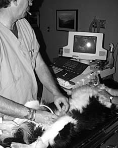
Fresh samples give the best results; samples should be analyzed within two hours, or up to six hours after collection if they have been refrigerated. Samples can be collected via catheter, cystocentesis (removal of urine by using a sterile needle to tap through the abdominal cavity into the bladder), or by catching a mid-stream flow in a clean container – easer said than done, especially with small dogs and females who squat low to the ground.
Each of these collection methods has its advantages and disadvantages, and often, unless you are quick with the catch-jar and quick afoot to deliver it to the vet clinic, it may be easiest to have the technicians collect it at the clinic and read it in-house. Depending on what condition is suspected, there may also be a best time of day for collection; check with your vet.
In the lab, the urine is observed for abnormalities of color or odor; specific gravity is determined by placing a drop of urine on a hand-held instrument called a refracto-meter; and various chemicals in the urine are analyzed by dipping a chemically-impregnated dip stick into the urine and observing color changes. Finally, the sample is centrifuged and the sediment is analyzed under the microscope to detect the presence of cells, casts, crystals, microorganisms, and tumor cells.
Note: Urine provides a welcome place for bacteria to grow; bacterial contamination from the surrounding environment is a common error when a sample is not handled properly or when it is left on the counter for hours before being analyzed.
When I did relief work in different clinics, I often saw stains that had become contaminated with bacteria or yeasts, leaving the clinic staff with the impression that all the urine samples run in their clinic came from infected animals. I made it a habit to put a little stain on a slide and look for bacterial contamination under the microscope before I ran any urine samples.
• Color and odor. Urine color is clear when dilute; it is normally yellowish due to the urochromes in the urine, and the yellow color intensifies when concentrated (i.e., when the animal is dehydrated). Urine can pick up a variety of colors and odors, and these may indicate disease, diet, or drugs. For example, a cloudy appearance may be due to urinary tract cells, bacteria, fat, crystals, or mucus; an examination of the sediment will differentiate among these possibilities. Red colored urine may be due to red blood cells, hemoglobin, recent ingestion of beets, or one of several drugs.
Urine with a strong smell of ammonia may have come from an infected urogenital tract; some bacteria are urea splitters, creating the smell of ammonia.
• Specific gravity. Specific gravity measures the density of the urine, relative to the mass of an equal volume of water, and it is determined by using a refractometer. Osmolality, a measure of the number of solute particles within the urine, may also be used to differentiate diseases.
One of first signs of renal tubular disease is the loss of the concentrating ability of the tubules. Normal canine specific gravity is usually more than 1.030. A specific gravity above or below 1.010 ± 0.002 indicates functional capacity.
A “fixed” specific gravity of 1.008 to 1.012 (isosthenuria) indicates that the tubules are not functioning normally. A specific gravity of less than 1.008 may indicate an early disease condition: diabetes insipidus, hypoadrenocorticism (Addison’s disease), or primary renal disease.
The key word in the above is “fixed.” For example, fluid therapy may temporarily lower the value below 1.008, but if the tubules are functional, the value will return to above 1.012 after therapy has ceased. The specific gravity of a dehydrated dog’s urine may be in excess of 1.030, but after rehydration, he should display more normal values.
While specific gravity is a key assay for determining kidney function, if a problem is suspected, it should always be performed along with tests to determine the dog’s blood urea nitrogen (BUN) and creatinine levels to diagnose or rule out renal failure.
“Dip Stick” Tests for Dogs
The following tests – pH, proteins, glucose, ketones, occult blood, and bilirubin – are performed by immersing a “dip stick” into fresh urine and observing color changes due to chemical reactions from the reagents contained in small patches located along the reagent stick.
Some dip sticks also contain reagents for nitrite or leukocyte esterase. A positive nitrite test indicates that bacteria may be present in significant numbers, especially Gram negative rods such as E. coli. Leukocyte esterase measures the presence of white blood cells, whether they are intact or lysed (partially destroyed or dissolved). Thus a positive test indicates infection; a negative test indicates that an infection is unlikely.
• pH. Urine pH is dependent on the animal’s diet. In herbivores it is alkaline; carnivores and omnivores have acid to alkaline urine, depending on the amount of protein in the diet.
Urine acidity may also be caused by starvation, fever, metabolic or respiratory acidosis, prolonged muscular exercise, or administration of acid salts (e.g., ammonium chloride). Urine alkalinity may be due to bacterial infections (cystitis), metabolic or respiratory alkalosis, or ingestion of sodium bicarbonate.
• Proteins. A small quantity of protein passes the glomerular filter but is reabsorbed by the renal tubules; consequently, normal urine is usually negative when tested for protein. In concentrated urine (specific gravity greater than 1.050) a reaction level ranging from trace to 1+ may be normal.
A slight transitory proteinurea may be associated with fever, muscular exercise, or seizures. A false positive may occur with alkaline urine (pH greater than 8.5), and either hemoglobin or myoglobin in the urine may also cause false positive results.
A consistent presence of more than a trace of protein in nonconcentrated urine indicates the need for further diagnostics to determine the cause. Possible causes include: inflammation of the lower urinary tract (or cystitis, which is often accompanied by the presence of urine-discoloring red or white blood cells); abnormalities to the filtration system (glomerulonephritis and renal amyloidosis are the most common causes); or possibly from prostatitis, urethritis, and vaginal or preputial discharges. Rarely, a form of tumor (plasma cell tumor) produces low molecular weight proteins (Bence Jones proteins) that pass through the kidney’s filter, and that may create a positive protein test.
• Glucose. The presence of urinary glucose is a primary screening test for diabetes mellitus. The normal dog’s kidney can reabsorb blood glucose amounts up to about 180 mg/dl, and only amounts over this value will be spilled into the urine.
The dip stick test for glucose should normally be negative. Diabetes mellitus is due to an absolute or a relative lack of insulin and is defined as persistently high glucose (greater than 180-200 mg/dl) in the blood.
There are several artifacts, depending on the type of reagent strip used, that may interfere with dip stick tests for glucose.
False negatives may be caused by refrigerated urine; large amounts of ascorbic acid from high levels of vitamin C or tetracycline therapy (the therapeutic vitamin C interference with test results is especially a consideration if your dog is on high doses of vitamin C); salicylates; or large amounts of protein in the urine.
Falsely increased values may be caused by hydrogen peroxide or bleach (caused by collecting the sample in an old bleach bottle, for example). Also, many antibiotics may cause false positive results.
While the most common cause of glucosuria is diabetes mellitus, there are other physical causes that may elevate blood sugar high enough to be read on the dip stick. Stress may cause a slight and transitory elevation (especially in cats), renal tract hemorrhage, renal tubular dysfunction, and hyperadrenocorticism (Cushing’s disease).
In addition, there are several breed-specific diseases that are not related to diabetes that cause glucose spillage into the urine. (This glucosuria-causing phenomena has two different etiologies and has been reported in the Basenji, Norwegian Elkhound, Shetland Sheepdog, Miniature Schnauzer, Scottish Terrier, and mixed breeds.)
A positive glucose test in the urine is an indication that a blood glucose test should be performed.
• Ketones. Excessive ketones are produced when the animal is metabolizing fatty acids as an energy source. Slight ketonuria can be seen in malnourished dogs, and it frequently accompanies advanced cases of canine diabetes mellitus.
The odor of ketones can be detected on the breath or in the urine of fasting/starving animals – and, incidently, on the breath of some people who are dieting. The ketone odor indicates the person’s body is metabolizing excess body fat. Some people can detect the odor readily – it smells like fingernail polish remover; others are not as sensitive.
• Occult blood. A positive reaction indicates red blood cells, free hemoglobin (from the breakdown of red blood cells), or myoglobin (a byproduct of muscle breakdown).
A positive reaction must be interpreted in light of what is seen in the sediment. Red blood cells or red cell casts may be seen in the sediment; their presence reflects hemorrhage in the urinary tract.
If RBCs are not seen, the positive reaction may be from hemoglobin, indicating that either the RBCs have broken up in the urine, releasing free hemoglobin, or the positive test may be due to myoglobin, the oxygen-transporting pigment of muscle.
• Bilirubin. Bilirubin is a pigment found in liver bile, and it is formed mainly from the breakdown of red cells and the subsequent release of the hemoglobin they contained.
Bilirubin appears in the urine if there is an increase in the serum concentration. Small amounts (trace to 1+) are normal, but a result of 3+ or higher is significant and indicates the need for an evaluation of the status of the red cells, possibly along with further liver-function tests.
Some Causes of Discolored Urine
| Urine Color | Possible Causes |
| Yellowish-orange, green or black | Bilirubin |
| Brown or rust-yellow | Metronidazole |
| Sulfonamides | |
| Red-brown | Myoglobin |
| RBCs | |
| Hemoglobin | |
| Dilantin | |
| Chronic lead or mercury intoxication | |
| Red-purple | Porphyrins |
| Phenolphthalein | |
| Red | RBCs |
| Hemoglobin or myoglobin | |
| Dyes | |
| Beets | |
| Blue | Methylene blue |
| Blue-green | Urinary tract infection due to Pseudomonas aeruginosa |
| Milky | Infection (pyuria) |
| Dietary fat |
Canine Urine Sediment Evaluation
After the urine has been evaluated visually and via the dip stick, the sample is centrifuged, the fluid portion discarded, and the remaining nonfluid and cellular elements evaluated under the microscope. Results are reported in the average number of cells or other elements observed in each high power field (/hpf) or low power field (/lpf). Normal urine collected by cystocentesis contains only small numbers of cells and other formed elements from the urinary tract.
• Leukocytes (white blood cells). Normal values may be 0 to 3/hpf in urine collected by cystocentesis. Increased numbers (pyuria) supports the presence of inflammation (cystitis).
• Erythrocytes (red blood cells). Normal urine may have a few red cells (0 to 3/hpf). Increased numbers indicate inflammation or hemorrhage. If the red cells are seen in a cast (see below), hemorrhage in the kidney is suggested. Blood in the urine (hematuria) may be associated with stones in the urinary system (uroliths), tumors, bacterial infection, trauma, sterile cystitis, a variety of kidney diseases, urinary parasites, and thrombocytopenia (decreased numbers of platelets or thrombocytes, the clot-forming cells in the blood).
• Cells. A few large and small round cells may appear in normal urine, but their numbers may be increased in animals with cystitis, tumors, or other inflammation of the urinary tract. Evaluation of the urine sediment under the microscope is a good way to screen for urinary tract tumors.
• Casts. Urine casts are cylindric molds of the kidney tubules, formed of aggregated proteins or cells within the tubules and then passed into the urine, where they can be seen on microscopic exam. Urine from normal animals contains only a few hyaline casts (2 or less/lpf) or granular (1 or less/lpf). The type of cast present represents a continuum of severity of disease – from mild (hyaline) to more severe (granular) to very severe (waxy). The causes and significance of urine casts are summarized in the box above.
• Bacteria. Bacteria may be introduced to normal urine through the collection process when catheterization or midstream collection are used. Bacteria found in urine collected by cystocentesis indicate an infectious process. If a significant bacterial infection is found, your veterinarian may order or perform a urine Gram stain test to identify the bacteria and determine the most appropriate antibiotic for treatment.
• Yeasts and fungi. These are contaminants, which may have been introduced during collection or through contaminated stains used to evaluate sediment.
• Crystals. Since the appearance of crystals in the urine (crystalluria) may be a normal finding, their presence needs to be evaluated against the pH and concentration of the urine. We look to the pH because some crystals will normally be seen in acid urine; others require an alkaline media to form. For example, triple phosphate crystals are associated with some urinary stone (calculi) formation, but are more often present in alkaline urine without the presence of calculi.
We also consider the concentration of the urine. Crystals detected in dilute urine are more significant than crystals seen in concentrated urine, where more crystals might be expected.
Certain crystals may be of diagnostic significance. For example: cystine crystals may be associated with cystine uroliths, ammonium biurate suggests liver insufficiency, and ethylene glycol (antifreeze) toxicity often creates a characteristic Maltese cross crystal.
• Sperm. Sperm are found in about one fourth of urine samples taken by cystocentesis from intact males and recently bred females. In unneutered males, a certain number of misguided sperm must swim through the vas deferens (the exit tube of the testes) via the ejaculatory duct, through the prostate into the urethra, and then into the bladder. In the female, errant sperm, splashed about the female’s vulva during breeding, must be able to swim via the urethra into her bladder.
Interpreting Urine Casts
| Type of Cast | Associated With | Interpretation |
| Hyaline | Protein in the urine | Insignificant |
| Epithelial | Kidney tube sloughing | Acute severe tubular damage |
| Cellular to granular | Tubular epithelial cell degeneration | Suggests tubular disease |
| Waxy | They develop from cellular and epithelial casts | Indicate a chronic tubular lesion |
| Leukocyte (white blood cell) | Kidney inflammation | Suggests kidney infection |
| Erythrocyte (red blood cell) | Hemorrhage | Usually the result of trauma |
Final Diagnosis from a Urinalysis
Although the urinalysis may be the most straightforward of the diagnostic tests available, there is still a touch of art-form in its interpretations. It is an inexpensive test, and almost every veterinary clinic can perform one in-house, although some clinic managers prefer to send them to commercial veterinary labs.
Finally, as always, the results of the urinalysis need to be correlated with other observations, the history of the dog, with other tests, and with the signs and symptoms the dog is demonstrating. In the end, though, the urinalysis is one of the most vital tools available for the diagnostician.
Dr. Randy Kidd earned his DVM degree from Ohio State University and his Ph.D. in Pathology/Clinical Pathology from Kansas State University. A past president of the American Holistic Veterinary Medical Association, he’s author of Dr. Kidd’s Guide to Herbal Dog Care, and Dr. Kidd’s Guide to Herbal Cat Care.


This article is an excellent resource. But I am curious, I have a 14 year old cocker spaniel and she marks a lot of territory but I can tell when she is urinating more – there IS more – and she drinks a lot of water. We did a lab test and found high lycotyte numbers – I wonder how to tell the if this is a bladder, UTI or kidney infection? Or does it matter in the short term?
As simple as this may sound, my pet died from complications from this procedure.
I had a Cocker mix who urinated on every blade of grass when we would go out for walks. She never had a UTI or problems with accidents in the house or getting up in the middle of the night. We just figured she was marking her territory. If she has a large increase in white blood you definitely should be asking more questions. I would compare the current labs from both vets to previous labs & I would start asking questions about other illnesses such as cancer could she have. I don’t know what the 2nd doctor suggested the problem was but I’d certainly be doing some research to find out what questions you need to ask. Considering her age & a “large increase in the number of white blood cells” the first question I would be asking is “is it cancer”. I question a doctor who is quick to suggest a UTI if the symptom is just frequent urination on walks…she could be doing it because it smells like a “bathroom” to her. It’s a little late now but I sure would look at her labs & compare to previous labs. …perhaps find a better vet who doesn’t jump to conclusions because she frequently piddles on walks but not at home. If your dog had a UTI, she would be frequently urinating at home, not just on walks where she smells all the placed other dogs had pee’d & figures it’s time to pee!
I’m currently going through issues with my 12 year old flat coat newfie mix who has dementia & is now asking to go out every hour & gets up in the middle of the night to go out….never piddles on walks &. I make a list of all symptoms, do some homework on reputable websites, compare labs & ask LOTS of questions of my vet.
This was very interesting! My dog,
A 10 year old Great Dane/Lab was diagnosed with a UTI. I had brought her to the vet about s month ago after observing very frequent urination on a morning walk. I was told she had a large increase in the number of white blood cells and needed to be on Amoxicillin. HOWEVER, she did not ask to go out frequently nor to ask to go out in the middle of the night. I took her to another vet to repeat the test. She found no indication of a UTI. I do notice she is doing more urination on walks and wonder if I should have more testing done.