Basic screening tests, in combination with regular physical exams, are foundation components of a good health care program. In younger dogs, routine tests are done to establish normal baselines, exclude congenital problems, and/or ensure safety for anesthesia. In older pets, these tests often provide the first indication of possible health problems.
Last month, we described some basic screening tests that veterinarians use to check for early signs of illness. The test results of senior dogs, in particular, are more likely to possess abnormalities, ranging from subtle and easily explained irregularities to complex abnormalities that require further work-up.
So what happens when the screening test shows a problem? Let’s explore the next-step diagnostics.
Abdominal Ultrasounds for Dogs
What it is: Ultrasound technology uses sound waves bounced off of structures to create a picture. When used in a medical sense, this tool can look at the structure of organs in the abdomen and chest or ligaments and tendons. Abdominal ultrasound shows the structure and internal texture of the abdominal organs, including the liver, gallbladder, kidneys, spleen, small and large intestine, bladder, adrenal glands, and lymph nodes among others.
Why run it: Unlike an x-ray, which can only show the outline of things, an ultrasound can show the internal structure and architecture. This is important, as a change in the texture of an organ can indicate disease. Ultrasound is also very useful in detecting masses. Frequently, masses do not cause any outward changes and can be missed on physical exam or even x-ray, but an ultrasound can pick up very small growths. This early detection allows for more options moving forward. A very large mass may not be surgically removable, whereas when detected early, when still small, surgery may result in a cure.
Ultrasound is also used to guide a veterinarian’s needle to obtain samples of organs for biopsy; without this guidance, surgery would be required to access the organs for a biopsy.
When it should be run: Abdominal ultrasound is a test that is recommended most often based on changes to lab work. For example, if a routine chemistry panel shows elevation in liver enzymes, an ultrasound can be used to evaluate why those values are elevated. Alternatively, if your veterinarian feels a suspicious area on physical exam, she may recommend an ultrasound to check for a mass or organ enlargement.

Case example: Roswell, a nine-year-old Golden Retriever, presented to his veterinarian for his annual physical. His owner mentioned that he had seemed lazier lately and wasn’t eating his food as quickly as he had when he was a younger dog. On his physical exam, Roswell’s gums were noted to be a little lighter in color than normal and his belly seemed uncomfortable when the veterinarian was checking it. A complete blood count and chemistry profile were recommended. These tests showed a low red blood cell count (anemia) and elevations in multiple liver values.
Based on these results, as well as the discomfort Roswell had shown during his physical and the comments of decreased energy and appetite, Roswell’s veterinarian recommended an abdominal ultrasound. The ultrasound revealed a liver mass that was slowly bleeding into Roswell’s abdomen.
Because it was detected early, Roswell was able to undergo surgery to have his tumor removed. While it was cancerous, it was completely removed and to date, almost a year after surgery, Roswell has had no further signs of illness and has regained his youthful spirit.
Echocardiograms for Dogs
What it is: An echocardiogram is an ultrasound of the heart. Similar to an abdominal ultrasound, an “echo” uses sound waves to create a picture of the heart.
An echocardiogram can provide a wealth of information about heart health. It can be used to evaluate the thickness of the heart muscle, the functionality of the valves, and the coordination of the beat. We can zero in on a single valve or look at the heart as a whole. We can evaluate the space around the heart for fluid or masses or look at how blood flows through the heart. Using a feature called a color Doppler, a veterinarian can assess the direction of blood flow across valves, into and out of the chambers of the heart, and through blood vessels.
Why run it: Characterizing heart disease is incredibly important to successful treatment. When your veterinarian can see exactly what is happening, appropriate medications can be prescribed to ward off heart failure or slow the progression of disease.
For example, a veterinary medication called pimobendan has been proven to prolong life when started in dogs with a heart condition called dilated cardiomyopathy (DCM) that are otherwise healthy. This is huge! Untreated, DCM can lead to congestive heart failure.
An echocardiogram also helps define severity of heart disease. This lets your veterinarian provide important information about prognosis and what to watch for.
When it should be run: The reason an echocardiogram is recommended depends upon the screening test that detected the possible abnormality in the first place. During a physical examination, your veterinarian may have heard a cardiac change that warrants further investigation. Detected with x-rays, an enlarged heart may have prompted the recommendation. Any time heart disease is suspected, an echocardiogram is the gold standard of diagnostics.
This test is especially important prior to undergoing an anesthetic procedure. Dogs with heart disease are at greater risk for complications from anesthesia, but these can largely be mitigated with an appropriate diagnosis and medical management. If your veterinarian recommends a “complete cardiac work-up,” an echocardiogram is the first step. Getting an exact diagnosis will allow your dog to live the longest, fullest life possible.
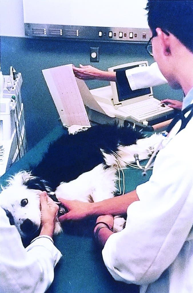
Electrocardiograms for Dogs
What it is: The electrocardiogram, also called an ECG or EKG, is a visual representation of the heartbeat. It transcribes the electrical impulse that causes your dog’s heart to lub-dub. There are three parts to an ECG – the P wave, the QRS complex, and the T wave. Each part represents a different portion of a single contraction. It is important that these all happen in a coordinated, predictable way to pump blood through the body effectively.
An ECG also measures heart rate and the spacing between beats. Interpreted all together, it creates a picture of your dog’s heartbeat.
Why run it: Abnormal heart rhythms cause myriad symptoms, from subtle things like general lethargy to more dramatic things like collapse. Certain breeds of dog are even prone to sudden cardiac death from abnormal heart rhythms. What you may see as a low drive to play ball may actually be weakness from a heart that isn’t beating right. There are medications to help manage these conditions and restore your pup’s normal energy level.

When it should be run: An electrocardiogram is part of a complete cardiac workup. In conjunction with an echocardiogram, it allows for complete assessment of heart health. Your dog’s heart, simply put, is what keeps him moving. If there’s a problem, it’s crucial to know quickly and get it under control. Dogs don’t have heart attacks the way people do, but they can die suddenly from untreated heart disease. Early detection is paramount to long-term management. Abnormal rhythms, independent of physical changes, can sometimes even be cured.
Case example: Sampson came in for his yearly exam and vaccinations. As part of his history, his owner mentioned that he had been having fainting spells about once every few weeks over the winter. He would be playing normally, then fall over and seem briefly unconscious. He always recovered within seconds and never seemed to have any lingering damage, so his owner didn’t think too much of the episodes.
During his visit, his veterinarian heard an abnormal heart rhythm and felt a racing pulse. Sampson’s owner approved an ECG, which showed him to be having runs of a very fast, abnormal heart rhythm intermixed with a normal heartbeat.
This finding prompted a visit to a cardiology specialist, who diagnosed Sampson with supraventricular tachycardia, a kind of fast heart rhythm that, if left untreated, can result in sudden death. The fainting spells seen by his owner happened when that rhythm occurred for too long without going back to normal, keeping blood from effectively getting to Sampson’s brain and other organs.
Sampson was started on medication and since that time has had no further fainting spells.
Complete Thyroid Panels
What it is: The thyroid gland secretes a number of different hormones that are responsible for regulating a multitude of things. Thyroid hormones have a hand in just about every process in the body. The complete thyroid panel measures thyroxine (T4), triiodothyronine (T3), and thyroid-stimulating hormone (cTSH). Having all of these values allows for a full evaluation of potential thyroid disease.
Why run it: There are two cases in dogs that result in low T4. The first is true hypothyroidism and the second is known as “sick euthyroidism.” When a low T4 value is noted on screening blood work, a complete thyroid panel is needed to differentiate between the two conditions. Dogs with true hypothyroidism require supplementation, whereas dogs with sick euthyroidism can worsen with supplementation.
When it should be run: We touched on this a bit in last month’s article. The time to run a complete thyroid panel is when the screening thyroid check comes back low. This allows your veterinarian to assess your dog’s need for supplemental thyroid hormone.
When a dog is truly hypothyroid, thyroid-stimulating hormone will be above normal limits. This is because the brain is trying to tell the thyroid gland to produce more hormone. A truly malfunctioning thyroid gland will not be able to increase production, leading to all thyroid hormones being low.
In the case of sick euthyroidism, thyroid-stimulating hormone will be low or normal, leading to low or normal thyroid hormones. Dogs who are sick will decrease thyroid hormone production naturally, but this is an appropriate response, so there is no need to start supplementation. Dogs who are truly hypothyroid need supplementation because their thyroid gland is not properly functioning.
These Tests Are Worth the Investment
There are so many ways that our dogs enrich our lives. They provide companionship when we are lonely, motivation to get out and exercise, and assistance in life and work, just to name a few things. We are entrusted with keeping them safe and keeping them healthy in return.
When your veterinarian recommends an advanced diagnostic test, such as the ones discussed here, her motivation is to get the most information possible to create a plan to prolong good quality of life. We all wish there was a crystal ball that would tell us what is wrong and a magic wand to wave and fix it. While we don’t have those things, we do have diagnostic tests!
After graduating from Michigan State University College of Veterinary Medicine in 2011, Kyle Grusling had internships in small-animal clinical medicine and surgery, then practiced emergency medicine for three years, before deciding to pursue a career in general practice at Northland Animal Hospital in Rockford, Michigan. When she’s not at work, Dr. Grusling enjoys spending time with her husband and their two sons, two cats, and Golden Retriever.


